---
language: "en"
---
# Mid Infrared Spectromicroscopy (Mid-IR) Beamline
*
## [Mid Infrared Spectromicroscopy (Mid-IR)](https://midir.lightsource.ca/mid-infrared-spectromicroscopy-mid-ir/home.md)
* [About Us](https://midir.lightsource.ca/mid-infrared-spectromicroscopy-mid-ir/about-us.md)
* [Software](https://midir.lightsource.ca/mid-infrared-spectromicroscopy-mid-ir/software.md)
* [Contact](https://midir.lightsource.ca/mid-infrared-spectromicroscopy-mid-ir/contact.md)
*
### [User Guide](https://midir.lightsource.ca/mid-infrared-spectromicroscopy-mid-ir/user-guide.md)
* [Reading](https://midir.lightsource.ca/mid-infrared-spectromicroscopy-mid-ir/reading.md)
* [FAQs](https://midir.lightsource.ca/mid-infrared-spectromicroscopy-mid-ir/faqs.md)
* [Before Beamtime](https://midir.lightsource.ca/mid-infrared-spectromicroscopy-mid-ir/before-beamtime.md)
* [During Beamtime](https://midir.lightsource.ca/mid-infrared-spectromicroscopy-mid-ir/during-beamtime.md)
* [After Beamtime](https://midir.lightsource.ca/mid-infrared-spectromicroscopy-mid-ir/after-beamtime.md)
*
### [News](https://midir.lightsource.ca/mid-infrared-spectromicroscopy-mid-ir/news.md)
* [Beamline News](https://midir.lightsource.ca/mid-infrared-spectromicroscopy-mid-ir/beamline-news.md)
* [Research Highlights](https://midir.lightsource.ca/mid-infrared-spectromicroscopy-mid-ir/research-highlights.md)
---
language: "en"
---
# About Us
## **Overview**
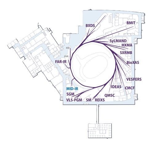
The Mid Infrared Spectromicroscopy beamline (Mid-IR) provides state-of-the-art Fourier Transform IR spectroscopy and microcroscopy. At Mid-IR we have three endstations that specialize in [++**synchrotron radiation spectromicroscopy**++](https://midir.lightsource.ca/mid-infrared-spectromicroscopy-mid-ir/bruker.md), [++**chemical infrared imaging**++](https://midir.lightsource.ca/mid-infrared-spectromicroscopy-mid-ir/agilent.md) and [++**time-resolved**++](https://midir.lightsource.ca/mid-infrared-spectromicroscopy-mid-ir/irsweep.md) measurements. Please check out each instrument page for more details.
The [**Bruker endstation**](https://midir.lightsource.ca/mid-infrared-spectromicroscopy-mid-ir/bruker.md) is optimized for high-brightness, diffraction-limited spectromicroscopy with synchrotron infrared radiation.
The [**Agilent endstation**](https://midir.lightsource.ca/mid-infrared-spectromicroscopy-mid-ir/agilent.md) is optimized for high spatial resolution, large field of view chemical infrared imaging.
The [**IRsweep endstation**](https://midir.lightsource.ca/mid-infrared-spectromicroscopy-mid-ir/irsweep.md) is optimized for high signal-to-noise, time-resolved spectroscopy measurements with microsecond time resolution.
## Our Mission
At Mid-IR we aim to provide state-of-the-art instruments to access high quality data and to build a national network of infrared researchers and users from coast-to-coast. We strive for excellent user support through all project stages including design, measurement and data analysis.
## What is infrared spectromicroscopy?
FTIR spectromicroscopy visually identifies the spatial distribution of specific chemical functionalities. The physics of long wavelength infrared light limits the spatial resolution, and the diffraction-limited spatial resolution is theoretically predicted to be 3 - 10 µm in the 4000 - 900 cm⁻¹ range.
A white light microscope is co-linear with the infrared microscope, and a visual image is collected of the sample. From this image, a feature or region of interest is selected. The infrared spectromicroscopy map or image is collected using an infrared sensitive detector. The resulting spectra can then be analyzed and used to identify the spatial distribution of chemical species or changes. To learn more, see [++further reading++](https://midir.lightsource.ca/mid-infrared-spectromicroscopy-mid-ir/reading.md).
---
language: "en"
---
# Acknowledgements
If you used Quasar or Orange-Spectroscopy for your data analysis, please cite us!
For more information, see
---
language: "en"
---
# Acknowledging the CLS
Researchers are asked to acknowledge the beamline(s), as well as the participation of any beamline staff who may have assisted in any aspect of an experiment, and include the following statement when presenting results from the CLS.
> Part or all of the research described in this paper was performed at the Canadian Light Source, a national research facility of the University of Saskatchewan, which is supported by the Canada Foundation for Innovation (CFI), the Natural Sciences and Engineering Research Council (NSERC), the National Research Council (NRC), the Canadian Institutes of Health Research (CIHR), the Government of Saskatchewan, and the University of Saskatchewan.
## Acknowledging Mail-in Results
For results obtain through the Mail-in Program, please include the acknowledgement above and acknowledge that data were collected by either:
* The person(s) who collected the data, if known, or
* Mid-IR staff
## Reporting Publications
Researchers are asked to [report any publications](https://www.lightsource.ca/users/your-cls-experiment/after-your-beamtime.php#ReportingPublications) based on work they performed, in whole or in part, at the Canadian Light Source. The list of the publications reported to the CLS is in important factor in our funding and is taken into account during peer review of research proposals.
Access the list of publications ([++https://user.lightsource.ca/pubs/++](https://user.lightsource.ca/pubs/)), click on the green \[ + \] button in the top right-hand corner of the page and follow the instructions to provide the requested information in the resulting dialog window. You must be a CLS user to submit a publication.
## Highlights!
If you'd like to be featured in [Mid-IR research highlights](https://midir.lightsource.ca/mid-infrared-spectromicroscopy-mid-ir/research-highlights.md), please [send us an email](https://midir.lightsource.ca/mid-infrared-spectromicroscopy-mid-ir/contact.md)with a figure/image, a summary and the citation of your paper!
---
language: "en"
---
# After Beamtime
## Yay data!
1. [Data Transfer](https://midir.lightsource.ca/mid-infrared-spectromicroscopy-mid-ir/data-transfer.md) to your personal/professional workstation.
2. Read the [++Quasar guide++](https://midir.lightsource.ca/mid-infrared-spectromicroscopy-mid-ir/software.md) to get started on your analysis.
3. Confirm samples have been dealt with appropriately (returned to user or disposed of)
4. [++Contact us++](https://midir.lightsource.ca/mid-infrared-spectromicroscopy-mid-ir/contact.md), we're here to help!
5. Communicate your results via publication, presentation and/or media. [Acknowledge](https://midir.lightsource.ca/mid-infrared-spectromicroscopy-mid-ir/acknowledging-the-cls.md) the CLS and the Mid-IR beamline!
---
language: "en"
---
# Agilent
## Agilent Cary 670 FTIR Interferometer with Cary 620 Microscope
### 🟢 Normal Operations
The Agilent Cary 670 spectrometer with a Cary 620 Microscope is a state of the art instrument for infrared spectromicroscopy and Chemical Infrared Imaging. The Cary FTIR system is equipped with a 128 x 128 pixel Focal Plane Array detector and provides high spatial resolution, large field of view, high sensitivity and fast collection times.
The system is ideal for imaging biomedical and biological materials, agricultural samples, materials such as polymers, art specimens, and other samples suited to infrared chemical analysis.
Advantages of the Agilent chemical imaging system include:
* large field of view (up to 2640 µm x 2640 µm in a single measurement)
* rapid acquisition times
* calibrated mosaic stitching for large-area measurements
* live FFT processing pipeline for as-collected, real-time data viewing and streamlined data processing
This spectrometer is an excellent complement to high-brightness synchrotron radiation FT-IR spectromicroscopy where rapid collection of large areas will help identify areas of interest for closer study.
### Schematic
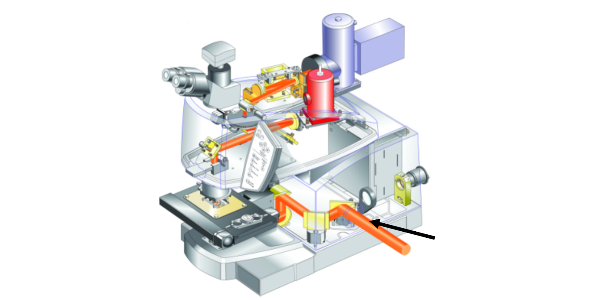
Agilent Cary 620 FTIR Microscope
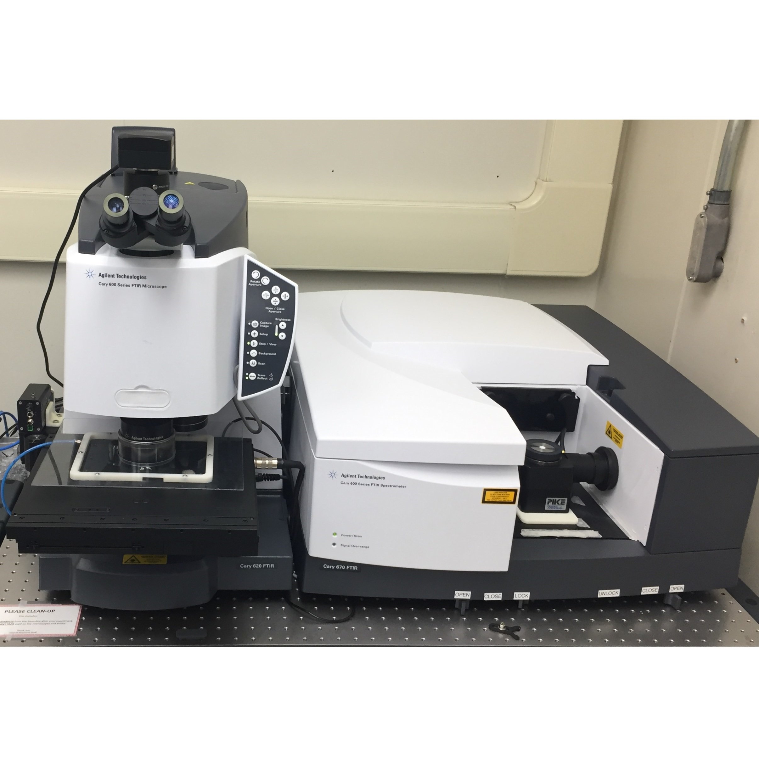
At the beamline
## Capabilities
|---------------------|------------|-----------------------------------------|-----------------------------------------|--------------------------------------|
| **Achieved Spatial Resolution Summary** |||||
| Objective, NA, mode | pixel size | achieved spatial resolution 3750 cm⁻¹\* | achieved spatial resolution 2500 cm⁻¹\* | Field of view (single FPA tile size) |
| 4x, standard | 20.6 µm | | | 2640 x 2640 µm |
| 15x, 0.62, standard | 5.5 µm | 6.9 µm | 7.6 µm | 700 x 700 µm |
| 15x, 0.62, high mag | 1.1 µm | 2.4 µm | 3.0 µm | 140 x 140 µm |
| 25x, 0.81, standard | 3.3 µm | 4.3 µm | 5.0 µm | 420 x 420 µm |
| 25x, 0.81, high mag | 0.66 µm | 1.4 µm | 1.7 µm | 85x85 µm |
\*actual achievable resolution will depend on sample and preparation, please consult beamline staff.
---
language: "en"
---
# Agilent 4X Image Overviews
## Return to [++Agilent Operation++](https://midir.lightsource.ca/mid-infrared-spectromicroscopy-mid-ir/agilent-operation.md) User Guide
The Agilent Wizard assists with sample location and experiment planning. At a minimum, you should ensure the objective setting on the *Define Regions* tab matches the infrared objective you plan to use.
*Important note:* The Agilent Wizard assists with experiment setup and planning, but **does not** affect the data acquisition step. All data acquisition is controlled through Resolutions Pro.
## Collect 4X Image Overviews of Each Sample
You can take 4X visible overview images of your sample(s) to assist with navigation when the purge shield is on. This will be done for you by beamline staff if you are operating remotely.
1. Ensure the 4X objective is in place - this step cannot be completed remotely.
2. Mount sample(s) on plate, and mount on microscope
3. In the Agilent Wizard header enter User name, Project, and Sample Name of first sample
4. Open *Imaging Method Editor* in Resolutions Pro:
1. Select Method "FPA Imaging/4X ZnSe Overview.clm"
2. *Visible Image*: find focus
3. With joystick, mark corners for visible mosaic and *Capture*
1. *Tip: If you are noting stage max/min positions while maneuvering around your sample to find corner positions,* ***only use the coordinates provided by the Wizard*** *as these will not be the same as on ResolutionsPro.*
4. View "Captured visible image"
1. Click somewhere on the image to make a single red rectangle (IR grid)
2. *Optional:* Using Windows Explorer, copy the 4X `VisMosaicCollectImages_Thumbnail.bmp` from `VisMosaicCollectImages` to Project folder
5. In the Agilent Wizard:
1. Click the "+" button next to "Visible image" list
2. Confirm ROI microscope setting
6. Repeat steps 3-5 for each additional mounted sample
1. In Wizard: Click "Add new Sample" and update sample name
7. In the Agilent Wizard, click the "Save" button.
Overview images can be restored in the case of computer restart using the "Restore" button.
8. Close Resolutions Pro *Imaging Editor* window when finished adding overview images
4x Objective
.png?cb=391a4db71942055d673b696fb9504aa1)
Set Corner 1 \& Corner 2 on opposite corners of each sample
*** ** * ** ***
## Begin Experimental Workflows
When transitioning from 4x overview image collection → IR measurements, remember to rotate the desired magnification objective in place and double-check that the condensor magnification matches the top and the purge shield is in place. The system is now ready to begin sample measurements.
### [Agilent Transmission Imaging Workflow](https://midir.lightsource.ca/mid-infrared-spectromicroscopy-mid-ir/agilent-transmission-imaging-workflow.md)
[Agilent ATR Imaging Workflow](https://midir.lightsource.ca/mid-infrared-spectromicroscopy-mid-ir/agilent-atr-imaging-workflow.md)
---
language: "en"
---
# Agilent ATR Imaging Workflow
## Return to [++Agilent Operation++](https://midir.lightsource.ca/mid-infrared-spectromicroscopy-mid-ir/agilent-operation.md) User Guide
This page is our User Guide for [++ATR Imaging++](https://midir.lightsource.ca/mid-infrared-spectromicroscopy-mid-ir/attenuated-total-reflection.md) with our [++Agilent++](https://midir.lightsource.ca/mid-infrared-spectromicroscopy-mid-ir/agilent.md) Endstation.
## 1. Load method file
In Resolutions Pro *Imaging Method Editor*, select your method file from the "Users" folder.
## 2. Find Centerburst
Press the "Find Centerburst" button.
* This will check operation of the interferometer and determine the current centerburst position.
* *Note this uses an internal detector and can be performed regardless of sample position in the microscope*
## 3. Focus sample
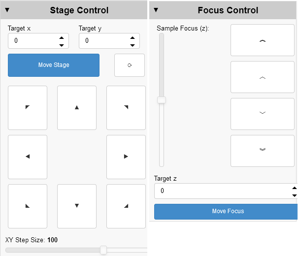
Sample position / focus can be controlled using the joystick or the Agilent Wizard *Stage Control* and *Focus Control* tabs.
* View the sample using "Visible Image"
* Switch to viewing the sample through the microscope eyepiece.
The spot that is in the cross-hairs of the eyepiece will be close to the center of the measured ATR region
*Tip*: If your samples are time/air sensitive, you can skip placing them on the stage now and wait until step 7. The background for the measurements is made with the Ge crystal in air, the sample does not influence the background.
## 4. Insert ATR crystal
Slide the Ge micro-ATR crystal assembly in place.
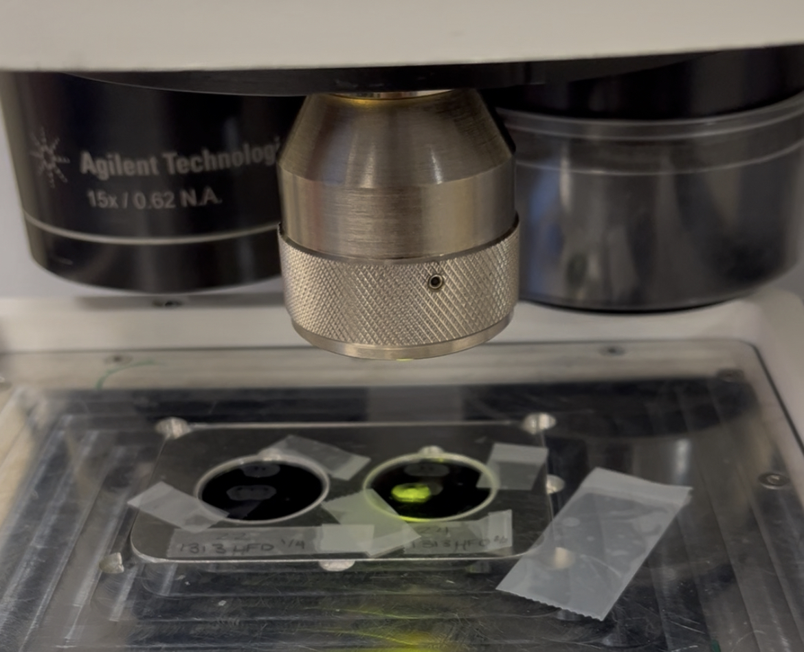
* The crystal will travel 1-2 mm above your sample at visible focus.
* **Germanium is brittle and easily scratched.** Check for interferences with this sliding motion.
## 5. Non-Uniformity Correction Calibration
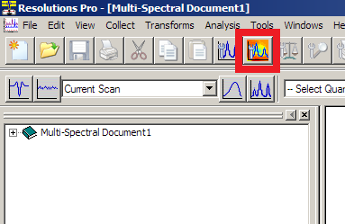
Open Resolutions Pro *Imaging Method Editor* "Live FPA" window
* Check that the FPA is at the operating temperature ( \<80 K)
* Select "Show Raw Data", "High Range"
* Adjust the vertical slider bar (integration time) so that the displayed signal is not greater than 70% of the full height.
*The 70% line is between the "Frame Rate" and "Frame Period" text lines.*
* Click "Calibrate" to perform the correction. The results of the correction will appear.
*You may wish to make a note of the integration time and resulting LowFlux/HighFlux values.*
## 6. Measure background
Click "Background" to start the background / reference measurement
* When the "Save as" dialog appears, navigate to the appropriate file location and then enter the **background** file name.
* Check the resulting IR image for appropriate signal levels or undesired material.
*Tip:* Do not re-use background file names.
## 7. Visible sample image
* Slide ATR crystal into visible light position
* Open Resolutions Pro *Imaging Method Editor* "Visible Image" tab.
* Confirm sample position and focus
* Click "Capture"
* Using Windows Explorer, copy the `VisMosaicCollectImages_Thumbnail.bmp` from `VisMosaicCollectImages` to sample folder
## 8. Approach sample
* Slide the Ge crystal assembly into the measurement position
* Click `Live FPA` and select `Show Calibrated data`
* Adjust the contrast to teal/green or green/yellow range
* Watch the live Lancer Calibration view and use the fine adjust knob on the joystick to slowly approach the sample (raise the stage)
* A change in the image indicates ATR contact with the sample.
* Move the stage very slowly upwards until there is contact across the measurement field of view
## 9. Start sample scan
Click `Scan` button and save in the same folder as the background and visible image.
## Next sample / New region / Increase pressure
After collection, review the spectra across the IR image.
If the absorbance values are low, there may not been sufficient contact with Ge crystal. Repeat from [Step 8. Approach sample](https://midir.lightsource.ca/mid-infrared-spectromicroscopy-mid-ir/agilent-atr-imaging-workflow.md#8.-Approach-sample) and gently approach another 50 z units (\~¼ turn of stage controller)
**Before moving to another region or sample, you must retract the crystal to avoid damage.**
Depending on your sample, you may need to remove the crystal assembly and gently clean with alcohol/water and lens paper.
Next Steps:
[Move to next region and focus](https://midir.lightsource.ca/mid-infrared-spectromicroscopy-mid-ir/agilent-atr-imaging-workflow.md#3.-Focus-sample)
[Take a new background measurement](https://midir.lightsource.ca/mid-infrared-spectromicroscopy-mid-ir/agilent-atr-imaging-workflow.md#6.-Measure-background)
[Collect visible image](https://midir.lightsource.ca/mid-infrared-spectromicroscopy-mid-ir/agilent-atr-imaging-workflow.md#7.-Visible-sample-image)
[Approach Sample](https://midir.lightsource.ca/mid-infrared-spectromicroscopy-mid-ir/agilent-atr-imaging-workflow.md#8.-Approach-sample)
[Measure next Scan](https://midir.lightsource.ca/mid-infrared-spectromicroscopy-mid-ir/agilent-atr-imaging-workflow.md#9.-Start-sample-scan)
If you see a central speckle pattern in the background image, you have too much light on the detector. You should repeat the calibration and lower the integration time slider.
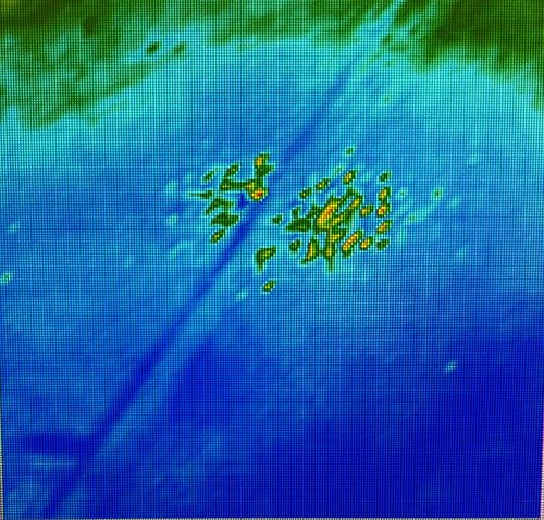
*Tip:*Travel distance will vary depending on the sample hardness, initial contact expected around 200-300 z units of travel
---
language: "en"
---
# Agilent Operation
## Agilent Instrument User Guide
[**About the Agilent**](https://midir.lightsource.ca/mid-infrared-spectromicroscopy-mid-ir/agilent.md)
[**Agilent 4X Image Overviews**](https://midir.lightsource.ca/mid-infrared-spectromicroscopy-mid-ir/agilent-4x-image-overviews.md)
[**Agilent Transmission Workflow**](https://midir.lightsource.ca/mid-infrared-spectromicroscopy-mid-ir/agilent-transmission-imaging-workflow.md)
[**Agilent ATR Imaging Workflow**](https://midir.lightsource.ca/mid-infrared-spectromicroscopy-mid-ir/agilent-atr-imaging-workflow.md)
## Instrument Orientation
### FPA Detector and Liquid Nitrogen
The FPA (imaging) detector is cryogenically cooled for operation with liquid nitrogen. A single detector fill will last for approximately 8 hours. It is very important **not to allow the detector to warm up** while it is powered on.
Liquid nitrogen filling is handled by a Norhof auto-filler, so you do not need to be present at the beamline to refill the detector. The auto-filler operates on a **4 hour** refill cycle. It is possible to trigger an early detector fill to accommodate your experimental schedule. Take note of detector temperature and check the status of the Norhof auto-filler at regular intervals during your experiment. At the end of your beamtime, beamline staff will direct you how to leave the detector / auto-filler. If in doubt, keep the auto-filler running.
### Spectrometer and Microscope
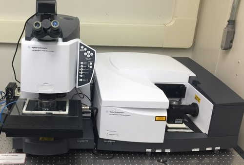
Cary 670 Spectrometer (right) and Cary 620 Microscope (left).
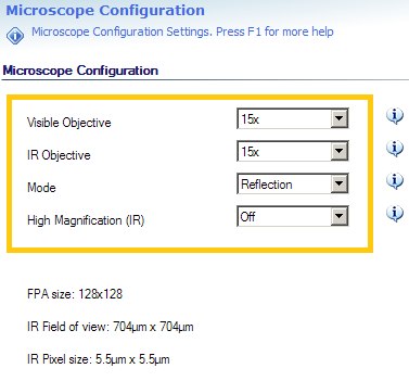
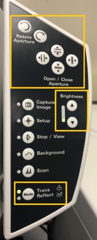
### Microscope controls
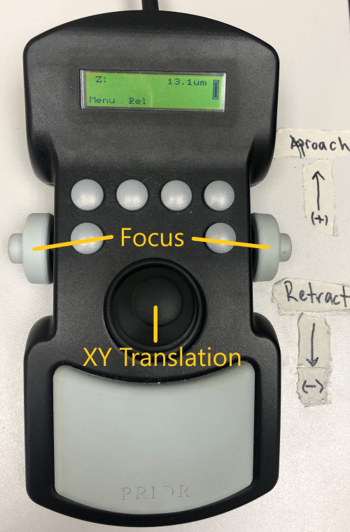
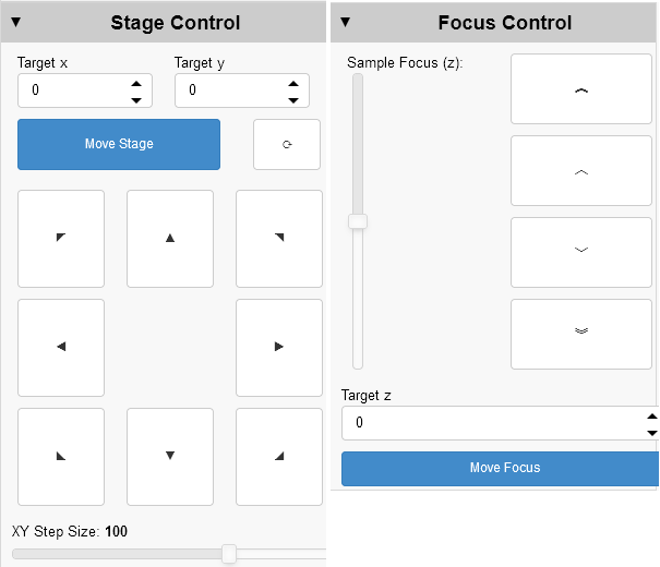
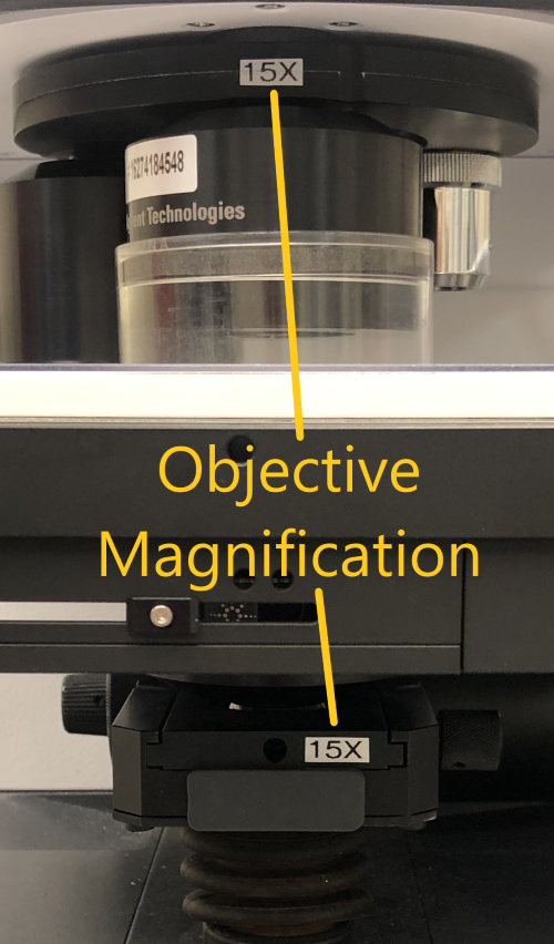
Manual and software controls exist for most microscope functions.
### Optics / Accessories
The Cary microscope is equipped with both 15X and 25X magnification objectives, as well as a 4X visible-only objective for sample viewing. There are internal zoom optics available which further increase the magnification by a factor of 5 at the cost of signal intensity at the detector. See the [Capabilities section](https://midir.lightsource.ca/mid-infrared-spectromicroscopy-mid-ir/agilent.md#Capabilities) of the [++Agilent++](https://midir.lightsource.ca/mid-infrared-spectromicroscopy-mid-ir/agilent.md) page for more details.
The 15X objective can be configured with a Ge [++ATR imaging++](https://midir.lightsource.ca/mid-infrared-spectromicroscopy-mid-ir/attenuated-total-reflection.md) crystal.
The main compartment of the Cary spectrometer can be configured for bulk spectroscopy using standard transmission mounts, a single-bounce Ge ATR or a multi-sample autosampler in both the Mid- and Near-IR.
The purge gas is constantly supplied to the sample but the purge shields must be in place. Ensure the clear plastic top plate and objective collar are in place for at least 15-20 minutes before acquiring data. This will provide adequate time for the purge gas to replace the ambient air in the system after the chamber is closed.
### Software
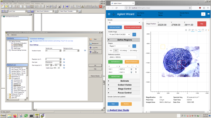
Resolutions Pro (left) is the primary software for instrument control and data acquisition.
The Agilent Wizard (right) software assists with stage control, sample orientation and experiment planning.
### Experimental Parameters
* Spectral resolution and objective magnification trade off measurement time per area and S/N with increased spectral/spatial resolution.
* Number of scans directly trades measurement time for S/N.
* Pixel aggregation (binning) trades file size and S/N for spatial resolution, with no impact on measurement time.
## Experimental Setup
These steps should be completed for you by beamline staff, however you should confirm the flap positions and detector cooling before starting your work. If you are remote, you can only confirm that the auto-filler is running.
1. Start the Norhof liquid nitrogen auto-filler to cool the FPA using the "Fill FPA" desktop link.
2. Open microscope input flap between FTIR and microscope.
3. Ensure there is a light path through the main FTIR compartment to the internal DTGS detector and that the compartment flaps are open. Any of the following configurations will work for imaging measurements:
1. Pike MIRacle ATR accessory
2. Multisample wheel (wheel removed)
3. Empty compartment (with purge cover)
4. Mode specific setup:
**Transmission** Confirm that the installed condenser matches the desired magnification
**ATR Imaging** Confirm the ATR crystal mount is installed and the FPA has been resized to 64x64 pixels
5. Turn on the detector
### Resolutions Pro Method Settings
Experimental settings are controlled through the Resolutions Pro Imaging Method Editor. Settings specific to your experiment can be saved to a custom method in the *Users* folder. After saving a new method, ***you should close and re-open the Imaging Method Editor window***.
Before starting your first measurement, **double-check the Imaging Method Editor Settings**.
Imaging Method Editor Settings
| **Page** | **Setting** | **Description** | **Comment** | **Remote Control** |
|-----------------------------------------------|---------------------------------|---------------------------------------------------------------|----------------------------------------------------------------------|--------------------|
| Microscope Configuration | Visible Objective | Objective used to collect Visible images | Setting must reflect objective in use | ❌ |
| | IR Objective | Objective used to collect IR hyperspectral images | Setting must reflect objective in use | ❌ |
| | Mode | Optical configuration, can be Transmission / Reflection / ATR | Changes microscope between Transmission / Reflection optical paths | ✅ |
| | High Magnification | Enable HiMag IR optics (additional 5X) | Adds / removes internal 5X optics from IR optical path | ✅ |
| Common Settings | Number of scans | Number of co-added scans | Background is commonly set to match sample | ✅ |
| | Resolution | Spectral resolution desired | Determines time to collect a single scan | ✅ |
| | Scan type | Y-axis output of calculated spectra (Absorbance/Reflectance) | | ✅ |
| | Scan range | Energy range to store in calculated output | Maximum range (FPA) is 3925 - 850 cm⁻¹, recommended 3900 -- 900 cm⁻¹ | ✅ |
| Advanced Settings: Collect | Speed | Interferometer mirror velocity | Set to 2.5 kHz for 128x128 FPA | ✅ |
| | Interferogram Sampling Interval | Data sampling interval | Set to 4 for 128x128 FPA | ✅ |
| | Number of Pixels Aggregated | Spatial averaging (binning) during collection | Influences S/N and file size, but not collection time | ✅ |
| Advanced Settings: Spectrometer Configuration | IR Source | | Set to "Rear: MIR Source" | ✅ |
| | Beam splitter | Set to "KBr" | Must reflect installed beam splitter | ❌ |
| | Aperture | | Set to "Open" for FPA Imaging | ✅ |
| | Beam Attenuator Throughput | | Set to "100%" for FPA Imaging | ✅ |
## Agilent Wizard
The Agilent Wizard assists with sample location and experiment planning. At a minimum, you should ensure the objective setting on the *Define Regions* tab matches the infrared objective you plan to use. *Important note:* The Agilent Wizard assists with experiment setup and planning, but **does not** affect the data acquisition step. All data acquisition is controlled through Resolutions Pro.
The Wizard program may be launched from the Agilent desktop shortcut. Visible orientation images are collected of samples loaded onto the stage mounts. Before starting the IR experiment, we will obtain 4x visible images of each sample and import them into the wizard following this procedure: [Agilent 4X Image Overviews](https://midir.lightsource.ca/mid-infrared-spectromicroscopy-mid-ir/agilent-4x-image-overviews.md). Once 4x images of all samples are collected, we can move on to experimental workflows.
## Experimental Workflows
The system is now ready to begin sample measurements.
* [Agilent 4X Image Overviews](https://midir.lightsource.ca/mid-infrared-spectromicroscopy-mid-ir/agilent-4x-image-overviews.md)
* [Agilent Transmission Imaging Workflow](https://midir.lightsource.ca/mid-infrared-spectromicroscopy-mid-ir/agilent-transmission-imaging-workflow.md)
* [Agilent ATR Imaging Workflow](https://midir.lightsource.ca/mid-infrared-spectromicroscopy-mid-ir/agilent-atr-imaging-workflow.md)
---
language: "en"
---
# Agilent Transmission Imaging Workflow
## Return to [++Agilent Operation++](https://midir.lightsource.ca/mid-infrared-spectromicroscopy-mid-ir/agilent-operation.md) User Guide
This page is our User Guide for [Transmission Imaging](https://midir.lightsource.ca/mid-infrared-spectromicroscopy-mid-ir/transmission.md) with our [Agilent](https://midir.lightsource.ca/mid-infrared-spectromicroscopy-mid-ir/agilent.md) Endstation.
## 1. Load method file
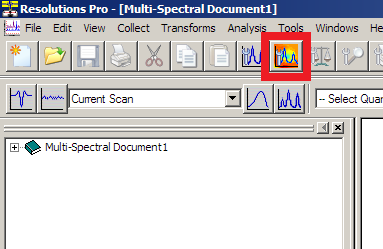
In Resolutions Pro *Imaging Method Editor* , select your method file from the "Users" folder.
🔑 This is a good place to double check that the IR objective lens and purge shield are correctly in place and the desired magnification is set up (4X, 15X or 25X).
## 2. Find Centerburst
Press the "Find Centerburst" button.
* This will check operation of the interferometer and determine the current centerburst position.
* *Note this uses an internal detector and can be performed regardless of sample position in the microscope*
## 3. Focus Sample
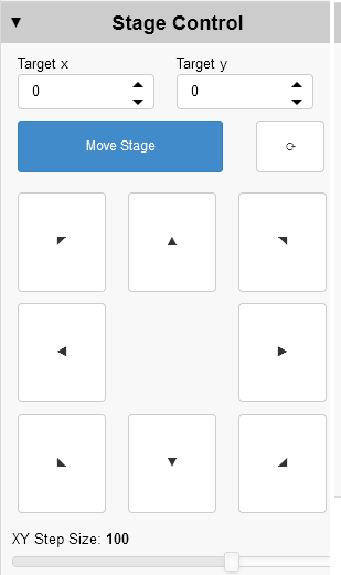
Sample position / focus can be controlled using the joystick or the Agilent Wizard *Stage Control* and *Focus Control* tabs.
* Move to the sample you wish to measure. View the sample using "Visible Image" tab in Resolutions Pro.
* Adjust the Stage Height (z) as needed to bring the sample into appropriate focus.
* You may need to adjust the sub-stage condenser to get a bright, even illumination.
* It is common practice to find the focus in reflection and then switch to transmission.
## 4. Mark sample Region of Interest
If you collected a 4X overview image of the sample,
* Select the Agilent Wizard "Edit ROI points" tool at the top of the figure.
* Click on the sample image to add ROI points
The ROI points will appear with estimated IR grid locations.
## 5. Locate clean background position
Using the joystick / Stage Control buttons, locate a position on your sample substrate which is clean from particles or contamination.
* Note that the IR field of view is larger than the visible camera.
* In Agilent Wizard, press "Set Reference" to store the background position
Use the Agilent Wizard to move to the sample. Add a ROI point -\> this will be added as the *last* point in the region. Make use of the "Move-To" feature to move the stage to this point.
Click on a coordinate to edit the ROI point
Click the **bin icon** to remove a row
Click the **numbers icon** to move the stage to that position
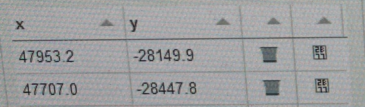
Agilent Wizard ROI Points
## 6. Non-Uniformity Correction Calibration
Open Resolutions Pro *Imaging Method Editor* `Live FPA` window. `Lancer Control` will open.
Look at the `Info` panel and check that the FPA is at the operating temperature:
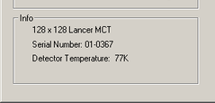
Detector Temperature \<80 K
* Select `Show Raw Data` in the `Calibration` box.
* (On-site) Adjust the sub-stage condenser to increase the signal while maintaining uniform illumination.
* Click `Calibrate` to perform the correction. The results of the correction will appear. *You may wish to make a note of the integration time and resulting LowFlux/HighFlux values.*
* Using ***Stage Control***, move the stage 100 μm ←/→ in X or ↑/↓ in Y and check the live image for deviations which indicate contamination on your reference position.

Adjust the vertical slider bar (controls the integration time) so that the displayed signal is not greater than 70% of the full height and/or the `Average Intensity` is \~9500. *The 70% line is between the "Frame Rate" and "Frame Period" text lines.*
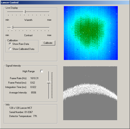
Show Raw Data
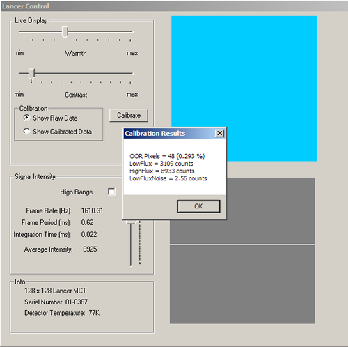
Calibration Result
If contaminants are observed, you must move to a new spot and recalibrate. [++Repeat from Step 6.++](https://midir.lightsource.ca/mid-infrared-spectromicroscopy-mid-ir/agilent-transmission-imaging-workflow.md#6.-Non-Uniformity-Correction-Calibration) Otherwise, return to the position where the current calibration was measured (Move to Reference) and proceed.
## 7. Measure background
Click "Background" to start the background / reference measurement. When the "Save as" dialog appears, navigate to the appropriate file location and then enter the unique background file name. For mosaic data collection, **create a folder with your sample name** and put the background file inside it. You can then use this folder as the mosaic save location and keep the background and sample files together. Give the background a distinct name, i.e. "...date_sample1_scan1_background" or similar.
Check the resulting IR image for appropriate signal levels or undesired material.
If you see a circular speckle pattern in the background image, the integration time is too long. You need to lower the integration time slider - you must **recalibrate** . [++Go back to Step 6++](https://midir.lightsource.ca/mid-infrared-spectromicroscopy-mid-ir/agilent-transmission-imaging-workflow.md#6.-Non-Uniformity-Correction-Calibration).
If you see evidence of sample or material in the background measurement you must **find a new background position** . [++Go back to Step 5.++](https://midir.lightsource.ca/mid-infrared-spectromicroscopy-mid-ir/agilent-transmission-imaging-workflow.md#5.-Locate-clean-background-position)
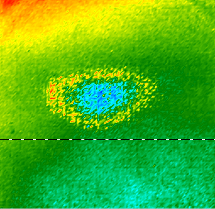
Improper background with speckle pattern
## 8. Visible sample image
In the Resolutions Pro *Imaging Method Editor* "Visible Image" tab:
* Select "Enable" in the "Visible Mosaic" section
* Open the Agilent Wizard Helper **Collect Visible** tab.
Press the Agilent Wizard Helper "Go ❶" orange button. The stage will move to the first corner of your region.
Press *Imaging Method Editor* "Set Corner 1" button
Press the Agilent Wizard Helper "Go ❷" orange button. The stage will move to the opposite corner of your region.
Press *Imaging Method Editor* "Set Corner 2" button
* Click *Imaging Method Editor* "Capture" to start visible mosaic collection. Open "Captured visible image" tab to monitor.
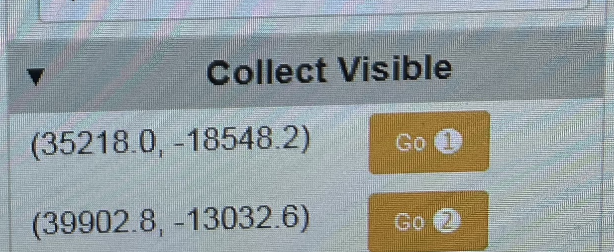
Agilent Wizard Go ❶ and Go ❷ Buttons
Visible Image Tip: For multiple tile mosaics the software will save the visible image when the IR sample data is collected. If you are only processing a single tile **you must save** the collected image to your project folder if you wish to use them. Use the `VisMosaicCollectImages` shortcut in Windows Explorer and save a copy to the folder with the matching IR data.
## 9. Define IR measurement area
In the Resolutions Pro *Imaging Method Editor* "Captured visible image" tab:
* Click and drag to define the desired measurement area. The locations to be measured will be drawn as red boxes.
* If the mosaic area is incorrect, select a new area
* If you wish, you can temporarily store this visible image in the Agilent Wizard with the "+" button next to the "Visible image" list.
Tip: The mosaic area always snaps to the top-left corner of the box drawn
**Before you start** - make sure you are confident with your sampling area and number of tiles to collect - once you proceed, you must commit to the measurement.
## 10. Start sample scan
Click the "Scan" button
* When the next dialog appears, navigate to your Project folder and
* (single tile) enter a unique file name in the Save as dialog
* (mosaic) select the folder you created when collecting the background
* Collection begins - you will see a dialogue box pop-up indicating the total number of tiles and measurement progress
Select a separate folder for each mosaic data collection - i.e. if you have to re-collect a scan, create a *new* folder for the new measurement.
At the end of the data collection, Resolutions Pro calculates the Fourier transform of the raw data. For a large mosaic, this can take 20-30 minutes, during which time the software will not provide any feedback.
*** ** * ** ***
## Next sample / region
Select your next sample/region in the Agilent Wizard and repeat as necessary.
[Move to the next sample and refocus](https://midir.lightsource.ca/mid-infrared-spectromicroscopy-mid-ir/agilent-transmission-imaging-workflow.md#3.-Focus-Sample)
[Mark your next sample ROI](https://midir.lightsource.ca/mid-infrared-spectromicroscopy-mid-ir/agilent-transmission-imaging-workflow.md#4.-Mark-sample-Region-of-Interest)
[Take a new background measurement](https://midir.lightsource.ca/mid-infrared-spectromicroscopy-mid-ir/agilent-transmission-imaging-workflow.md#7.-Measure-background)
[Collect visible image](https://midir.lightsource.ca/mid-infrared-spectromicroscopy-mid-ir/agilent-transmission-imaging-workflow.md#8.-Visible-sample-image)
[Start IR measurement](https://midir.lightsource.ca/mid-infrared-spectromicroscopy-mid-ir/agilent-transmission-imaging-workflow.md#10.-Start-sample-scan)
---
language: "en"
---
# Attenuated Total Reflection
## Background
In an Attenuated Total Reflection (ATR) measurement, the internal reflection element (IRE) is pressed in contact with the sample. An evanescent wave penetrates the surface layer of the sample to a depth of between 0.3 and 3 µm as a function of the measured energy. The constant penetration depth limits the effective pathlength through the material and allows measurement of thick samples with
### Samples
ATR is great for non-reflective samples and samples that cannot be thinly prepared (for transmission experiments). Direct contact between the germanium crystal and the sample is required and this may not be appropriate or desired in some cases.
### Results
ATR spectra are similar to transmission Absorbance spectra and can be qualitatively directly compared. Quantitatively, the absorbance reported varies as a function of energy due to changing penetration depth, which can be corrected with a simple calculation.
In the case of ATR imaging, the field of view is fixed and limited. Unlike other imaging methods, the images cannot be stitched together to create larger effective areas.
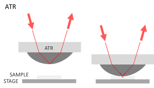
### **Available Accessories at Mid-IR**
Measurements which are possible depend on the ATR accessory used:
| **Accessory** | **Endstation** | **Measurement Type** | **Source** | **Internal Reflection Element** |
|---------------------------|--------------------------------------------------------------|------------------------------------------------------|--------------------|---------------------------------|
| Micro-ATR | [Bruker](https://midir.lightsource.ca/mid-infrared-spectromicroscopy-mid-ir/bruker.md) | single point spectromicroscopy | Synchrotron globar | Ge |
| Macro-ATR Imaging | [Bruker](https://midir.lightsource.ca/mid-infrared-spectromicroscopy-mid-ir/bruker.md) | ATR imaging (200 x 200 µm FOV) | Synchrotron globar | Ge |
| horizontal ATR microscope | [Bruker](https://midir.lightsource.ca/mid-infrared-spectromicroscopy-mid-ir/bruker.md) | custom ATR imaging \& single point spectromicroscopy | Synchrotron globar | User-supplied |
| ATR Imaging | [Agilent](https://midir.lightsource.ca/mid-infrared-spectromicroscopy-mid-ir/agilent.md) | infrared chemical imaging (70 x 70 µm FOV) | globar | Ge |
| Bulk ATR (PIKE) | [Agilent](https://midir.lightsource.ca/mid-infrared-spectromicroscopy-mid-ir/agilent.md) | bulk ATR | globar | Ge |
| Variable-angle ATR (PIKE) | [IRsweep](https://midir.lightsource.ca/mid-infrared-spectromicroscopy-mid-ir/irsweep.md) | time-resolved spectroscopy | globar ir laser | User-supplied |
---
language: "en"
---
# Beamline News
Beamline News
## Call for Proposals is Open!
### Scheduling Perion Jan-June 2027
The call for proposals is OPEN for beamtime in Cycle 43, January to June 2027. The call closes on Aug 19, 2026 at NOON Saskatoon time (CST).
Beamline contact:[**E-mail Scott Rosendahl, Mid-IR Beamline Responsible**](mailto:scott.rosendahl@lightsource.ca)
Learn more about [**Applying for Beamtime**](https://www.lightsource.ca/users/getting-started/applying-for-beamtime.php)and check out this[**Proposal Submission Webinar**](https://www.youtube.com/watch?v=q-M57H41xUg)video prepared by the USO.
Beamline News
## Cycle 42 is About to Begin!
### Beamtime occurring July-Dec 2026
Projects that have been allocated beamtime are being scheduled now. Stay tuned for scheduling notifications and updates from the USO and Beamline Responsible!
Use the [**User Portal**](https://user.lightsource.ca/) to check on your projects and scheduled beamtime.
Scheduling and beamtime request contact:[**E-mail Scott Rosendahl, Mid-IR Beamline Responsible**](mailto:scott.rosendahl@lightsource.ca)
Learn more about [**Applying for Beamtime**](https://www.lightsource.ca/users/getting-started/applying-for-beamtime.php)
*** ** * ** ***
Beamline News
## 2026 Annual Users' Meeting (AUM)
### May 21st, 2026 **at the University of Saskatchewan Campus / Virtual**
This one-day hybrid event will bring users, staff, and partners together to celebrate the rich and diverse science enabled by the CLS, and the researchers behind that work. In-person activities will be held in the University of Saskatchewan's Biology Building, Lecture Hall W.P. Thompson 106, with all sessions live-streamed for virtual participants. Everyone is welcome to join!
The AUM is **free to attend**, but you must register.
**For More Information:** [2026 CLS Annual Users' Meeting](https://event.fourwaves.com/cls-aum-2026/pages)
Registration is CLOSED as of 2026-05-07
## Beamline News
## Proposal Writing/Submitting Tips
[++Contact us!++](https://midir.lightsource.ca/mid-infrared-spectromicroscopy-mid-ir/contact.md) We can help you determine project feasibility, timelines and offer guidance in the proposal submission process. Our [User Guide](https://midir.lightsource.ca/mid-infrared-spectromicroscopy-mid-ir/user-guide.md) hosts more information about applying for beamtime and more! Check out our [About Us](https://midir.lightsource.ca/mid-infrared-spectromicroscopy-mid-ir/about-us.md) page for why our beamline is suitable for your experiment and samples.
**Mid-IR User Guide:** [Before Beamtime](https://midir.lightsource.ca/mid-infrared-spectromicroscopy-mid-ir/before-beamtime.md)
**CLS Guide:** [++Applying for Beamtime++](https://www.lightsource.ca/users/getting-started/applying-for-beamtime.php)
**Video Tutorial:** [++How to Submit a Proposal++](https://www.lightsource.ca/users/getting-started/user-portal-guide.php#HowtoSubmitaProposal)
## Beamline News
## Recent Publications
Yay data! Publishing research articles? Submitting a thesis? Attending conferences? Send us an [email](https://midir.lightsource.ca/mid-infrared-spectromicroscopy-mid-ir/contact.md)! Include a figure and summary of the work and we'll highlight it in our research news.
Make sure you [report your published works](https://www.lightsource.ca/users/your-cls-experiment/after-your-beamtime.php#ReportingPublications) to the CLS, too!
---
language: "en"
---
# Before Beamtime
## Planning and Coordinating Projects
Users wanting to submit new projects, ammend active projects or request some additional measurement time
1. Read our [Preparing a Proposal](https://midir.lightsource.ca/mid-infrared-spectromicroscopy-mid-ir/preparing-a-proposal.md) guide for creating and submitting proposals in the CLS User Portal
2. Monitor submitted proposals for Review and Clarifications
3. Ammend proposals to keep them current with changes to your samples or adjustments to the experimental plans
4. Keep in touch - you will be notified about call results and scheduling opportunities
**Notes about samples:** Significant sample amendments to a proposal should be discussed with the beamline responsible *one month* before beam time. **Any samples/materials/equipment that you plan on bringing must be listed on the project.** Samples/materials must be approved before arriving at CLS. To amend samples/materials/equipment, click the *"Amend"* icon under the "*Samples \& Materials"* section on the project page.
Amendments require review and approval prior to being used. **You will not be able to sign on to your beamtime session with Unapproved or In-Progress amendments. Please allow 2-4 weeks for reviews of Permit Amendments prior to the scheduled experiment.** Amendments submitted within two weeks of beamtime are not guaranteed to be processed and only the last valid samples and materials can be used for the experiment.
## Normal (on-site) Access
Users travel to the CLS to collect data in person.
1. Talk to beamline staff about your samples and setup requirements. [Contact](https://midir.lightsource.ca/mid-infrared-spectromicroscopy-mid-ir/contact.md) us!
2. Prepare for [++Facility Access++](https://www.lightsource.ca/users/your-cls-experiment/practical-information/facility-access-and-training.php) and be sure to complete [++Facility Access Training++](https://www.lightsource.ca/users/your-cls-experiment/practical-information/facility-access-and-training.php#FacilityAccessTraining) before your scheduled beamtime.
3. Review the [++Before your Beamtime++](https://www.lightsource.ca/users/your-cls-experiment/before-your-beamtime.php) and [++What to Expect On-Site++](https://www.lightsource.ca/users/your-cls-experiment/practical-information/what-to-expect-on-site.php) pages
## Mail-in and remote access
1. Talk to beamline staff about your samples and setup requirements. [Contact](https://midir.lightsource.ca/mid-infrared-spectromicroscopy-mid-ir/contact.md) us!
2. Prepare your [++shipment++](https://midir.lightsource.ca/mid-infrared-spectromicroscopy-mid-ir/shipping.md)
3. Ship samples close to beamtime
4. Contact beamline staff to confirm safe arrival of samples, verify storage methods and experimental plan
---
language: "en"
---
# Bruker
## Bruker Vertex 70v Interferometer with Hyperion 3000 IR Microscope
### 🟢 Normal Operations
The Bruker endstation provides a state-of-the-art Fourier Transform infrared spectrometer and microscope to supply diffraction-limited spatial resolution to an ever-widening range of infrared spectroscopy experiments.
This system is ideal for experiments that require the best available broadband brightness, signal to noise and spatial resolution.
Research and development to explore new experiments and re-examine existing techniques by applying the advantages of high brightness infrared synchrotron light are encouraged and actively pursued by beamline staff.
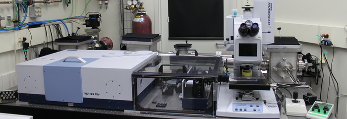
Bruker Endstation at the Mid-IR Beamline: Vertex 70v FTIR Spectrometer (left) and Hyperion 3000 Microscope (right)
### Techniques
[**Transmission**](https://midir.lightsource.ca/mid-infrared-spectromicroscopy-mid-ir/transmission.md)
[**Reflection/Transflection**](https://midir.lightsource.ca/mid-infrared-spectromicroscopy-mid-ir/reflection-transflection.md)
**Micro-** [**Attenuated Total Reflection**](https://midir.lightsource.ca/mid-infrared-spectromicroscopy-mid-ir/attenuated-total-reflection.md)
**Macro-Attenuated Total Reflection**
**Micro-Polarization Modulation Infrared Linear Dichroism**
### Detectors
**Single-element 100 µm MCT**
[**Focal Plane Array**](https://midir.lightsource.ca/mid-infrared-spectromicroscopy-mid-ir/focal-plane-array.md)**(64 x 64)**
---
language: "en"
---
# Bruker Operation
## Bruker Instrument User Guide
[**About the Bruker**](https://midir.lightsource.ca/mid-infrared-spectromicroscopy-mid-ir/bruker.md)
[**Bruker Transmission Workflow**](https://midir.lightsource.ca/mid-infrared-spectromicroscopy-mid-ir/bruker-operation-transmission-mapping-workflow.md)
[**Bruker Macro-ATR Workflow**](https://midir.lightsource.ca/mid-infrared-spectromicroscopy-mid-ir/bruker-operation-macro-atr.md)
[**hATR Microscope**](https://midir.lightsource.ca/mid-infrared-spectromicroscopy-mid-ir/hatr-microscope-operation.md)
## Instrument Orientation
### Detectors and Liquid Nitrogen
Both the single point mapping (MCT) and imaging (FPA) detectors are cryogenically cooled for operation with liquid nitrogen. A single detector fill will last at least **12 hours** for the MCT and up to a maximum of **8 hours** for the FPA.
It is very important **not to allow the FPA detector to warm up** while is it powered on.
The MCT should be allowed to thermalize for **20-30 minutes** after filling.
The detector will be initially filled with liquid nitrogen for you by beamline staff, however you will need to plan your experiments to accommodate refilling the detector.
Appropriate **PPE** is available at the beamline and must be worn whenever handling liquid nitrogen. If you are unsure how to fill the detector or have never done it, ask a member of the beamline staff to demonstrate. You should also have completed the **cryogenics safety training** module.
### Spectrometer and Microscope
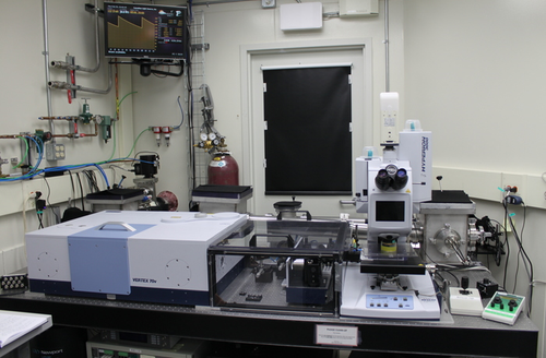
The instrument consists of a spectrometer (Vertex 70v, on left), the transfer optics (plexiglas box in center), and the Hyperion 3000 microscope (right). Synchrotron radiation enters the spectrometer from the rear out of the active optics chicane.
### Microscope Controls
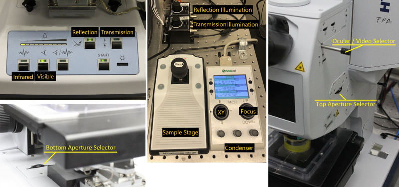
The Hyperion microscope can be controlled with buttons and knobs on the front/side. Top and bottom aperture wheel are rotated to select.
## Experimental Setup
These steps should be completed for you by beamline staff, however, you should confirm the detector cooling before starting your work.
1. Fill the detector(s) you will use for your experiments.
2. Confirm the installed objectives match the desired magnification.
3. Start OPUS using the icon on the desktop.
4. Configure Transfer Optics for experiment.
### Load Sample
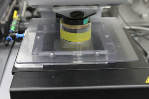
The purge system on the microscope stage reduces spectral interference from infrared-absorbing atmospheric gases such as water and CO2. Sample loading procedure:
* Mount your sample onto a sample plate. Use tape to secure sample disc to the plate.
* Remove the plexiglas purge components and rotate the top objective out of the way.
* Place your sample onto the microscope stage and return the objective to the working position.
* Reassemble the purge system around your sample.
You may find it helpful to mark key stage positions in the OPUS Video Wizard before closing the purge system, as it can be hard to see the sample position once the purge cover is in place.
### Video Wizard
In OPUS, open the Video Wizard using the "Start video wizard" button.
This will launch the microscope wizard and bring you to the Select Device window. Refer to the following Experimental Workflows to determine the correct entry for your experiment.
## Experimental Workflows
The system is now ready to begin sample measurements.
* [Bruker Operation \| Transmission Mapping Workflow](https://midir.lightsource.ca/mid-infrared-spectromicroscopy-mid-ir/bruker-operation-transmission-mapping-workflow.md)
* [Bruker Operation \| Macro-ATR](https://midir.lightsource.ca/mid-infrared-spectromicroscopy-mid-ir/bruker-operation-macro-atr.md)
---
language: "en"
---
# Bruker Operation | Macro-ATR
## Return to [Bruker Operation](https://midir.lightsource.ca/mid-infrared-spectromicroscopy-mid-ir/bruker-operation.md) User Guide
### Before You Start: Macro-ATR Component Orientation
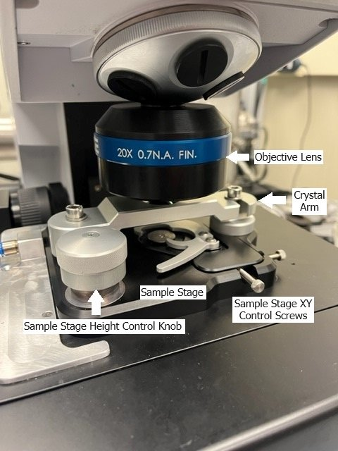
The Germanium single crystal ATR element is very fragile and expensive. You should **never** touch the crystal. Only clean with alcohols/water and only contact the crystal with appropriate lens paper for cleaning. When your sample is in contact with the crystal **do not** adjust the sample stage position - this could scratch the crystal.
There are *two* stages in this procedure, the sample stage and the microscopy stage. Make sure you are comfortable identifying them and orient yourself with how to control each.
* The **sample stage** height is controlled by rotating a screw located on the sample stage.
* The **microscopy stage** height is controlled by either the software *or* the black focus knob on the white box sitting to the right of the microscope.
If the microscope stage is moved too far forward it will fall off the rails and need to be reset by the beamline staff.
*This is avoidable* - **do not** move the stage unnecessarily far away from the sample location. **Rotate the objective lens** out of the way to give yourself a bit more room to change samples instead of driving the stage away from the lens.
## Macro-ATR User Workflow
### 1. Start the OPUS Microscopy Imaging Wizard
Start the OPUS Video Wizard
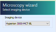
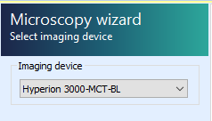
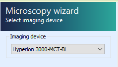
Select the `Hyperion 3000-MCT-BL` imaging device
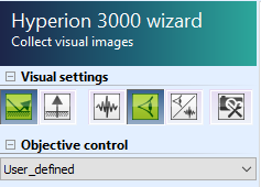
Select/confirm "User_defined" objective
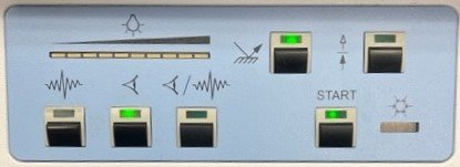
Select/confirm Hyperion is in *visible* and *reflection modes*
### 2. Define Measurement Location
Loading a sample is easiest with the objective rotated out of the way and the crystal arm swung out to give you more space.
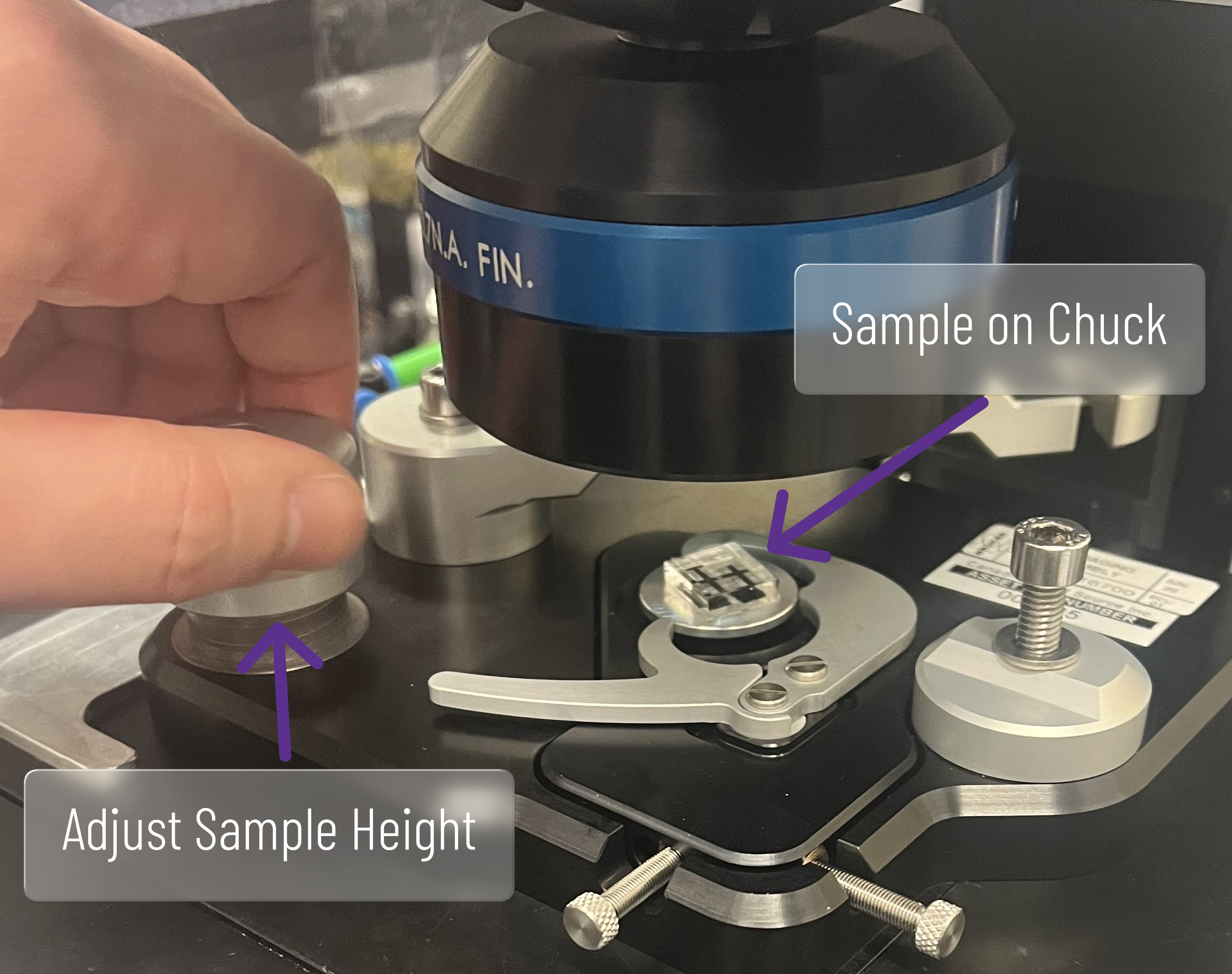
* Place chuck with sample onto macro-ATR sample stage. Move macro-ATR sample stage xy screws until chuck is reasonably centered and roughly adjust sample height using the **macro-ATR sample stage** z knob
* Rotate the objective lens back into place and open top aperture
* Move microscopy stage to center: in stage control select ⭐ and select "Zenith_DATE"
* Lower/raise **microscopy stage** z until sample is in focus
**COLLISION DANGER:** Ensure the crystal arm **does NOT collide** with objective when raising the stage.
* Situate your desired sample location directly below the objective and centered on the imaging location. Use macro-ATR sample stage xy screws until sample are of interest is visible and adjust microscope stage z as needed for visible focus.
### 3. Collect Visible Orientation Image
* Press "Define overview image". If no boundary points are set → load boundary points file and press "collect defined image"
* Once collection has finished, move stage back to center: in stage control select ⭐ and select "Zenith_DATE"
* Examine your image overview. If not satisfied with sample position repeat from [Step 2. Define Measurement Location](https://midir.lightsource.ca/mid-infrared-spectromicroscopy-mid-ir/bruker-operation-macro-atr.md#2.-Define-Measurement-Location)
Once satisfied with sample location and visible image, you must lower the sample stage completely before proceeding to avoid collisions.
Rotate the macro-ATR sample stage z knob to lower the sample all of the way.
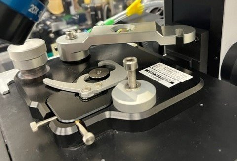
### 4. Focus SIR Light
Find SIR focus with the MCT detector.
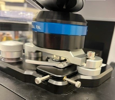
* Once sample stage is lowered, loosen screws of crystal arm
* Swing crystal arm into measurement position and tighten screws
**Make sure the crystal does NOT come into contact with the sample.**
**Make sure the washer is positioned between the arm and the screw head.** See image (right).
* Calibrate approximate microscope stage z-position for focus with SIR
* Raise microscope stage z for visible focus
**COLLISION DANGER:** Beware during raising of microscope z that crystal arm **does NOT collide** with objective
**Visible focus** should be near z=0. Visible light reflection will disappear and re-appear once close to focus. Rough focus is close when aperture is in visible focus when viewed through microscope ocular.
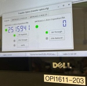
* Change Hyperion to IR mode. → Confirm Transfer Optics are in SR position (touchscreen OPI1611-203)
* Open advanced data collection and check signal tab. → Raise/lower microscopy stage z until counts is maximized (💡\>20,000 counts). Rotate the microscopy stage z focus knob only slightly. It is very sensitive. → Change aperture to desired size and optimize z until count is maximized
Counts will increase up to a maximum when you are at IR focus. If you overshoot the maximum point, lower the stage *slowly* until the raw detector signal is at a maximum.
Aim for: 0.3 mm aperture \~ 4,000 - 5,000 counts; 0.45 mm aperture \~ 8,000 - 9,000 counts.
* Set microscope stage z=0 ("Set to zero").
From this time point on, **do NOT change the microscope z position anymore** until finished with all IR measurements at this sample location
### 5. Take Background for SIR
* In stage control select ⭐ and select "Zenith_DATE" (stage should already be at this position)
* In video wizard, click "next" → "measure background once" → "measure at current position" → "measure background"
* Once complete, click "point at current location" **(complete at least once).**Note, this button looks like crosshairs.
* Check number of scans and wavenumber resolution → click "next"
* Provide sample name. E.g. "sample_SIR_bg" → once complete, click "next" → flat line IR spectra should be visible
### 6. FPA Background Setup and Calibration
Setup background collection parameters in the video wizard.
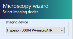
* Go back and select `Hyperion 3000-FPA-macroATR` imaging device
* Change Transfer optics to FPA (touchscreen OPI)
* **Open top aperture**
* In stage control select ⭐ and select "Zenith_DATE" (stage should already be at this position)
* Click "capture image" → Image should show up left (overview)
* Click "next" → "measure background once" → "measure at current position"
Calibrate FPA field flattening. A bright field image is obtained with all the light entering the detector while a dark field image is obtained with the light blocked from entering the detector.
* In stage control select ⭐ and select "Zenith_DATE" (stage should already be at this position)
* Select "live spectrum" → a flat illumination should be seen
* Right-click right bottom display and select "Customize Focal Plane Array settings..." → Check that FPA settings are set to **Offset 255 / Gain 1 / 32.47 µs**
* Calibrate FPA Camera
* Deselect the "FPA calibration" checkmark
* Click "acquire bright field image"
* Click "block IR beam" and while detector response is low, click "acquire dark field image"
* Select the "FPA calibration" checkmark and verify
* Click ok → click abort.*Note: check whether it goes to next tab*
### 7. Take Background for FPA
Measure the background for the FPA detector.
* Click "measure background". Check that **16 scans** will be collected (fewer scans are viable depending on sample)
* Once complete, mark a single measurement tile at the Zenith position → yellow box → Click "next"
* Provide sample name. *E.g.* "sample_FPA_bg" → click measure
* Once complete, click "next" → 2D hyperspectral image with flat line IR spectra at each pixel should be visible
Return to video wizard and return to Zenith position, select ⭐ and "Zenith_DATE"
### 8. Approach Sample with FPA
We need to make contact with the sample in order to view any spectral information. Raise the sample stage while monitoring the FPA signal until contact is achieved.
* Setup live view of FPA
* In video wizard, click "define overview" → "collect defined image"
* In stage control, select ⭐ and select "Zenith_DATE"
* Once complete, click "next" → "use existing background"
* Click "live spectrum"
* While observing the left display in live FPA, raise the sample stage until contact is made → click "Abort" to leave live FPA view once satisfied.
### 9. Measure FPA Overview
Setup FPA overview collection measurement.
* Right-click the left image tile image and select "load measurement position absolute" → load "`FPA_overview_grid`" file
* Check measurement is set to desired number of scans (suggested: **16** or fewer scans are viable depending on sample)
* Click "next" → give sample file name, e.g. "sample_FPA_overview_contact-01" → click measure → once completed, click "next"
Inspect the resulting IR image for areas of interest - use Orange workflow to do this.
**IF** no sample is visible or contact with sample in overview is not sufficient, increase pressure between sample and crystal by further raise the sample stage z: repeat procedure from [++8. Approach Sample with FPA++](https://midir.lightsource.ca/mid-infrared-spectromicroscopy-mid-ir/bruker-operation-macro-atr.md#8.-Approach-Sample-with-FPA).
**IF** an area of interest can be found, continue with following SIR measurement by proceeding to [++Step 10++](https://midir.lightsource.ca/mid-infrared-spectromicroscopy-mid-ir/bruker-operation-macro-atr.md#10.-Measure-SIR-Point-by-Point-at-Sample-Area-of-Interest). Otherwise skip to [++change sample section++](https://midir.lightsource.ca/mid-infrared-spectromicroscopy-mid-ir/bruker-operation-macro-atr.md#2.-Define-Measurement-Location) and attempt measurement with fresh sample.
Contact is made when 1) a steady change in colour for large areas can be observed in the FPA live view, 2) significant resistance can be felt at the sample raising knob and/or 3) the red limit line at the screw element is visible.
### 10. Measure SIR Point-by-Point at Sample Area of Interest
* Setup SIR point-by-point collection
* In video wizard, go back and select "`Hyperion 3000-MCT-BL`"
* Change Transfer Optics to SR (touchscreen OPI)
* Click "collect single image" - OR defined image to better see the measurement grid
* In stage control select ⭐ and select "Zenith_DATE"
* Once complete, click "next" → "use existing background"
* **Change top aperture** to either 0.3 or 0.45 based on experiment
* Setup measurement area
* Place points/line/grid measurement based on location in FPA overview image (from Quasar selection script)
* Check Signal
* Measure SIR
* Click "next" → give file name, e.g. "sample_SIR_region-01_contact-01" → click "measure" → once complete, click "next" and inspect measured IR data
* If a second area of interest is need, repeat steps from Click "collect single image" in [++Step 10.++](https://midir.lightsource.ca/mid-infrared-spectromicroscopy-mid-ir/bruker-operation-macro-atr.md#10.-Measure-SIR-Point-by-Point-at-Sample-Area-of-Interest)
* If completed measurement in this sample return to Zenith position (In stage control select ⭐ and select "Zenith_DATE"). Follow sample swap and clean up procedure**i**
* [Macro ATR Workflow Sample Change Procedure](https://midir.lightsource.ca/mid-infrared-spectromicroscopy-mid-ir/macro-atr-workflow-sample-change-procedure.md)
---
language: "en"
---
# Bruker Operation | Transmission Mapping Workflow
## Return to [++Bruker Operation++](https://midir.lightsource.ca/mid-infrared-spectromicroscopy-mid-ir/bruker-operation.md) User Guide
## 1. Select Device
In the OPUS Microscopy Wizard, select the **Hyperion 3000-MCT-BL** imaging device. This may have already been done during experimental setup.
If prompted to calibrate the stage, you must ensure there are no collisions. This entails:
* Remove the purge cover assembly
* If 36X condenser is installed, drop the condenser 6-7 mm.
## 2. Focus sample
Instrument with default to Reflection mode. Switch to Transmission.
Using either the video display or the ocular viewer, bring your sample into focus by twisting the sample stage joystick.
* Rotate joystick clockwise to lower sample stage, counter-clockwise to raise.
* Both apertures should be in the OPEN position
* Microscope should be in the visible light mode.
* Illumination is controlled by LED driver (intensity) and condenser stage joystick (alignment)
## 3. Collect visible image
* Ensure that `mode` and `objective` settings are correct (Transmission, 36x Objective for example):
If you are only interested in the part of the sample already in the field of view, press `Single image`. However if (more likely) you wish to survey a larger area of your sample, choose `Define overview image`.
* Navigate around the sample and press "Add new border point" at the edges of the area of interest
* Press "Collect defined image"
Multiple sample positions / visible images can be collected, but they must all be in the same plane of focus.
* The visible image(s) will be saved inside your OPUS data file.
* Right-click and select "Export current image" to save as stand-alone image files.
## 4. Locate clean background position
Continue through the OPUS wizard
* Select "Measure background once" or "Measure background after each n sample measurements" as desired.
* Select "User defined background position"
Using the joystick or saved stage position button (⭐), locate a position on your sample substrate which is clean from particles or contamination.
* Select "Set background position"
* Right click on the camera view and select "Add stage position" to store this position for subsequent measurements.
## 5. Align microscope
* Select the bottom aperture which corresponds to the desired spot size.
* Adjust the substage condenser position using the SmarACT controller to center and focus the aperture
* Select the matching top aperture
* Adjust the condenser position in XY to align the two apertures concentrically.
## 6. Measure background
Confirm microscope settings again (Transmission, Objective Magnification).
Verify signal through microscope:
* Switch to IR mode
* Press the "Advanced Data collection" button in the top toolbar
* Check the experiment file matches your method: `HYPERION 3000-MCT-BL_TRANS.XPM`
* Reload the correct file if different or if first time through wizard
* Switch to the "Check Signal" tab
* Select "Spectrum" view and verify energy curve and amplitude are nominal for your optical setup
* Beamline staff will assist with this during setup / training
* Close the "Advanced Data Collection" window
Collect your background by selecting "Measure background"
## 7. Define IR measurement positions
* Confirm desired number of scans under Measurement parameters / Scan time.
There are four types of measurement positions. They can be combined in any order or number.
To define a measurement position, select the desired tool and click on the visible image where you would like to measure.
1. **Mapping grid**
Click and drag to define a grid.
In the popup window, enter the desired point spacing and number of points, and press OK.
Reposition the grid if desired.
2. **Arbitrary points**
Each click will correspond to a single measurement.
Double-click the tool to select multiple points in a row
3. **Line scan**
Click and drag to define a line.
In the popup window, enter the desired point spacing and number of points, and press OK.
Reposition the line if desired
4. **Current stage position**
Pressing this tool will immediately mark the current stage position for measurement.
This can be combined with stage translation (Joystick or "Move to" stage tool) to mark multiple positions directly.
When all measurement positions have been selected, press "Next"
## 8. Start sample scan
* Enter a sample name
* Edit sample form if desired
* For example, enter selected aperture size
* Confirm file path corresponds to the current active project
* Press "Measure sample"
## Next sample / region
After collection, another visible image window will appear. This can be used to collect visible images after measurement if desired.
* Press "Next"
* Review data in OPUS if desired, or unload file and sync to review in Quasar
* Select "Video Guided Measurement" tab to return to Microscopy wizard
* Continue at step [++3. Collect visible image++](https://midir.lightsource.ca/mid-infrared-spectromicroscopy-mid-ir/bruker-operation-transmission-mapping-workflow.md#3.-Collect-visible-image)
---
language: "en"
---
# Contact
|  | **[++Scott Rosendahl++](mailto:Scott.Rosendahl@lightsource.ca)** Senior Scientist and Beamline Responsible, Mid-IR Room 2039 306-657-3667 |
| -20260507-224901.JPG?cb=4dd70029570d1c0695bbc9b1d18658f4) | **[++Grace Flaman++](mailto:Grace.Flaman@LIGHTSOURCE.CA)** Associate Scientist, Mid-IR Room 2039 306-657-3735 |
|  | **[++Amanda Quirk++](mailto:Amanda.Quirk@LIGHTSOURCE.CA)** Scientist, Bio/Life Science and Mid-IR Room 2074 306-657-3755 |
|  | **[++Kaiyang Tu++](mailto:Kaiyang.Tu@LIGHTSOURCE.CA)** Scientist, Strategic Support Group and Mid-IR Room 2079 306-657-3710 |
|---------------------------------------------------------------------------------------------------------------------------------------------------------------------------------------------------------------------------------|-------------------------------------------------------------------------------------------------------------------------------------------|
### Local Emergency Contacts
* Floor Coordinator: **306-657-3639**
* Available whenever there is stored current
* Emergency: **911**
* U of S Security: **306-966-5555**
* CLSI HSE
* 24/7 On-call: **306-227-3113**
* Business Hours: **306-657-3663**
### Beamline Phone Numbers
* **306-657-3607** - Beamline and User area
* **306-657-3615** - User area alternate
* **306-657-3586** - Room 1070 (IRsweep)
### Mailing Address
Canadian Light Source Inc.
University of Saskatchewan
44 Innovation Boulevard
Saskatoon, SK
Canada S7N 2V3
* **Reception:** **306-657-3500**
* **Fax number:** **306-657-3535**
### [++**CLS Contacts and Staff Directory**++](https://www.lightsource.ca/about/contact.php)
---
language: "en"
---
# Custom Code
You may already have some custom algorithms implemented in Python which have not ([++yet!++](https://midir.lightsource.ca/mid-infrared-spectromicroscopy-mid-ir/development.md)) been integrated as a widget in Quasar / Orange. This section will give an introduction to the **Python Script** widget and some details when integrating with spectral data in Quasar.
This page assumes familiarity with the Scientific Python ecosystem and with Python programming.
## Python Script widget
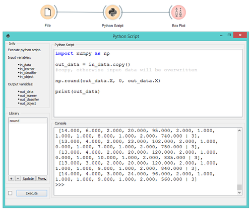
The Python Script widget provides programmatic access to your data in the middle of a workflow. This can be either pre-scripted (*Python Script* pane) or interactive (IPython *Console* pane). Scripts can be saved as stand-alone `*.py` files and loaded into new workflows or shared with your colleagues.
An excellent introduction to this widget for general use in Orange can be found in the Orange widget catalog: [++Orange Data Mining - Widget Catalog++](https://orangedatamining.com/widget-catalog/)
## Orange Scripting
A full introduction to scripting Orange is beyond the scope of this page. The following resources can get you started:
* [++Orange Data Mining Library: Tutorial++](https://orange-data-mining-library.readthedocs.io/en/latest/)
* [++Orange Data Mining Library: Reference++](https://orange-data-mining-library.readthedocs.io/en/latest/#reference)
In particular, the [++Data model++](https://orange-data-mining-library.readthedocs.io/en/latest/reference/data.html) and [++Data Table++](https://orange-data-mining-library.readthedocs.io/en/latest/reference/data.table.html) pages will help orient you to the `Orange.data.Table` data structure.
*** ** * ** ***
## Quasar Spectral Data
If you are used to working with spectral data in [++NumPy++](https://numpy.org/) or [++pandas++](https://pandas.pydata.org/) data structures in Python, you may want to skip learning the Orange Table data structure and quickly get your data in a familiar object. The following sections will point you how to access the data and navigate some of the special structure in Quasar.
### Energy Axis
The energy axis (wavenumbers, eV, etc) is stored as column headers (`mydata.domain.attributes`). To build a 1D array of energies from a spectroscopy Table, use the built-in `getx()` function on your table:
* `energy = orangecontrib.spectroscopy.data.getx(mydata)`
Note that if any attributes cannot be converted to `float`, the function will simply return an array of indices.
### Spectral Data
The spectral data itself is stored in a 2D numpy array accessible at `mydata.X`.
Spectra are stored row-wise, and normal numpy slicing can be used:
* `mydata.X[2]` would return the spectrum in the 3rd row of the table.
### Hyperspectral Cube
To reshape the hyperspectral data from a flatten 2D representation in the spectroscopy Table to a cube array, you need to provide the meta attributes where the x and y coordinate axes are stored. For most hyperspectral datasets, these are labelled *map_x* and *map_y* . This example results in the hypercube accessible as `hypercube` and the energy, x and y axes as `energy`, `map_x`, and `map_y`, respectively.
`import numpy as np`
`from orangecontrib.spectroscopy.data import getx`
`from orangecontrib.spectroscopy.utils import get_hypercube`
`mydata = in_data.copy()`
`energy = getx(mydata)`
`x_attr = mydata.domain["map_x"]`
`y_attr = mydata.domain["map_y"]`
`hypercube, lsx, lsy = get_hypercube(mydata, x_attr, y_attr)`
`map_x = np.linspace(*lsx)`
`map_y = np.linspace(*lsy)`
### Output your result
#### Existing data Table:
If you have just in-place modified your (copied) data, you can simply output the modified table directly:
* `out_data = mydata`
Remember not to in-place modify the original `in_data` object, always use `mydata = in_data.copy()` first.
#### Create new data Table:
If you have a new data set with no connection to the previous, you can create a new Table. This will break any Domain / Instances connection to the input data.
* `out_data = orangecontrib.spectroscopy.data.build_spec_table(features, data, additional_table=None)`
where
* `features` is a 1D numpy array defining the energy axis (wavenumbers, eV, etc) (size m)
* `data` is a 2D numpy array (shape (n, m)) with values
* (optional) `additional_table` is an Orange.data.Table with only meta or class attributes (size n)
### pandas DataFrame
Orange has built-in helpers for converting to/from `Orange.data.Table` and `pandas.DataFrame` data structures. This may be useful if you are already comfortable working with pandas.
* `df = Orange.data.pandas_compat.table_to_frame(in_data)`
* `table = Orange.data.pandas_compat.table_from_frame(df)`
Note this is not perfectly lossless: in particular, metadata axes important for hyperspectral data may be lost.
---
language: "en"
---
# Custom Figures
Plotting infrared spectra and hyperspectra can be fun, but frustrating! This tutorial is a starting place to make the your plots and figures look the way you want without leaving Quasar/Orange.
There are many resources that go further than this tutorial. Check out: and
## Matplotlib tutorial
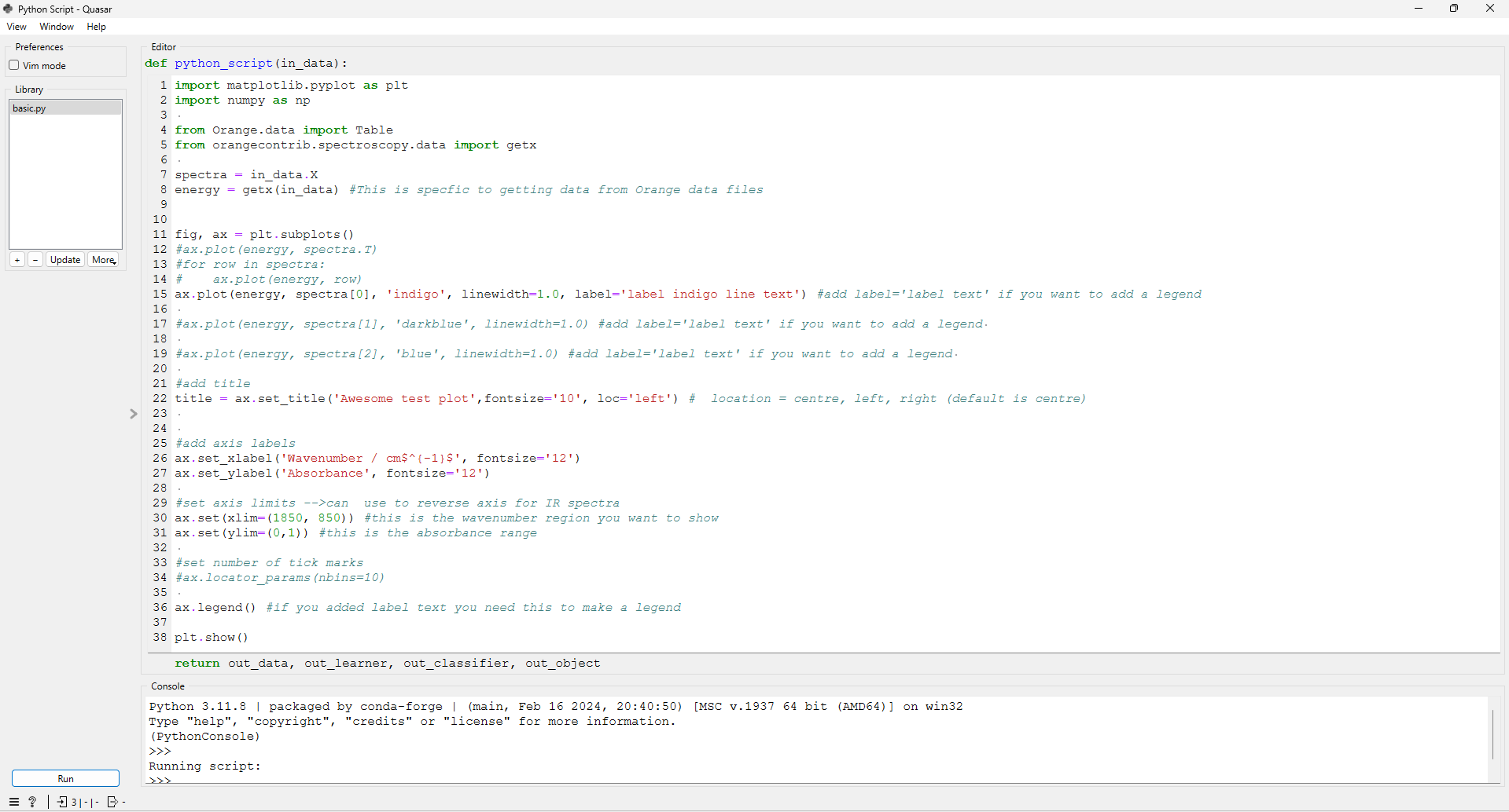
In Quasar, you can use the "python script" widget to format plots using matplotlib.
Python Script Widget
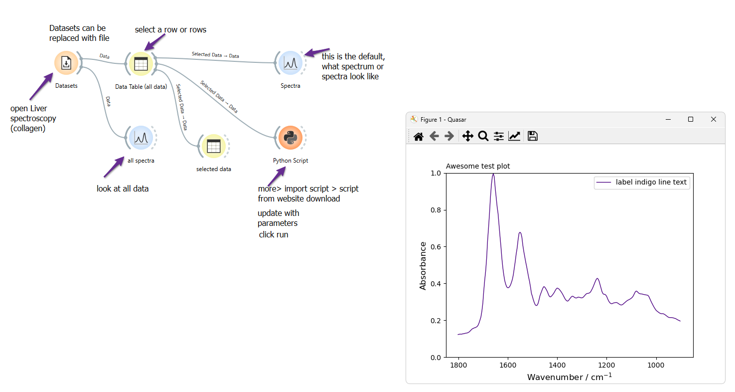
This workflow shows the process starting with data from quasar datasets. The same sequence could be used by selecting spectra from any data table, spectra plots or hyperspectra. Attach the python script widget to the output of your selected data, and format your plots from the script window.
[++Matplotlib tutorial++](https://midir.lightsource.ca/__attachments/a_96f850b6bcc4f5a6d3de93559de62fd369ccca0105a9cd9cdd3ca22ca5fa357a/matplotlib_tutorial.ows.md?cb=a0c89caca81d5b0abe3c10484437020f)
*** ** * ** ***
## Basic Scripts
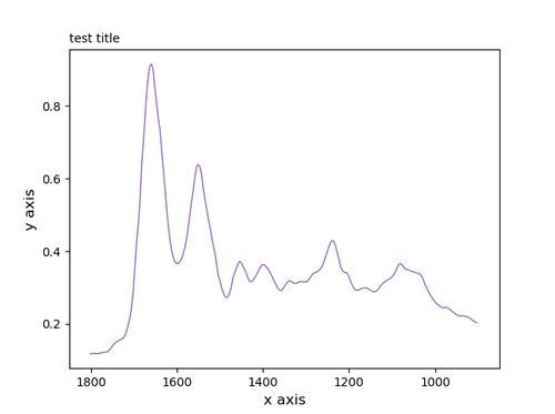
Below are a series of figures, click on the expand and copy the text into the Python Script widget to generate the plot in Quasar.
### Python Script: One Curve, One Plot
Python Script: One Curve, One Plot
import matplotlib.pyplot as plt
import numpy as np
from Orange.data import Table
from orangecontrib.spectroscopy.data import getx
spectra = in_data.X
energy = getx(in_data) #This is specific to getting data from Orange data files
###############################################################
fig, ax = plt.subplots()
###############################################################
ax.plot(energy, spectra[0], 'indigo', linewidth=1.0, label='label indigo line text')
#add label='label text' if you want to add a legend
#choose colour
#choose line width
###############################################################
#add title
title = ax.set_title('Awesome test plot',fontsize='10', loc='left') # location = centre, left, right (default is centre)
###############################################################
#add axis labels
ax.set_xlabel('Wavenumber / cm$^{-1}$', fontsize='12')
ax.set_ylabel('Absorbance', fontsize='12')
###############################################################
#set axis limits -->can use to reverse axis for IR spectra
ax.set(xlim=(1850, 850)) #this is the wavenumber region you want to show
ax.set(ylim=(0,1)) #this is the absorbance range
###############################################################
#add legend
ax.legend() #if you added label text use this to make a legend
###############################################################
plt.show()
*** ** * ** ***
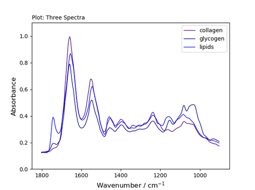
### [Python Script: Three Curves, One Plot with Legend](https://midir.lightsource.ca/__attachments/a_32339b79af0d3235b7340bb653d91f6c589852019eb9abfbd324d22a5e093fca/basic_1-3.py.md?cb=f1fbce3d16a73b36086546f3067f6226)
Python Script: Three Curves, One Plot with Legend
import matplotlib.pyplot as plt
import numpy as np
from Orange.data import Table
from orangecontrib.spectroscopy.data import getx
spectra = in_data.X
energy = getx(in_data) #This is specfic to getting data from Orange data files
fig, ax = plt.subplots()
################################################################################
ax.plot(energy, spectra[0], 'indigo', linewidth=1.0, label='collagen') #add label='label text' if you want to add a legend
ax.plot(energy, spectra[1], 'darkblue', linewidth=1.0, label='glycogen') #add label='label text' if you want to add a legend
ax.plot(energy, spectra[2], 'blue', linewidth=1.0, label='lipids') #add label='label text' if you want to add a legend
################################################################################
#add title
title = ax.set_title('Plot: Three Spectra',fontsize='10', loc='left') # location = centre, left, right (default is centre)
################################################################################
#add axis labels
ax.set_xlabel('Wavenumber / cm$^{-1}$', fontsize='12')
ax.set_ylabel('Absorbance', fontsize='12')
################################################################################
#set axis limits -->can use to reverse axis for IR spectra
ax.set(xlim=(1850, 850)) #this is the wavenumber region you want to show
ax.set(ylim=(0,1.1)) #this is the absorbance range
################################################################################
ax.legend() #if you added label text you need this to make a legend
################################################################################
plt.show() #show plot
*** ** * ** ***
### [Python Script: Two Plots, Three Curves](https://midir.lightsource.ca/__attachments/a_4093c4c71a8305639dc178fc54241d35ce02400f78e9d7d488f23e1a063f3eb0/basic2-3.py.md?cb=9119d98a58ddf8dc12d0b21eaa0e8a94)
Python Script: Two Plots, Three Curves
import matplotlib.pyplot as plt
import numpy as np
from Orange.data import Table
from orangecontrib.spectroscopy.data import getx
spectra = in_data.X
energy = getx(in_data) #This is specfic to getting data from Orange data files
################################################################################
fig, (ax, ax2) = plt.subplots(1,2)
################################################################################
ax.plot(energy, spectra[0], 'indigo', linewidth=1.0, label='collagen') #add label='label text' if you want to add a legend
ax.plot(energy, spectra[1], 'darkblue', linewidth=1.0, label='glycogen') #add label='label text' if you want to add a legend
ax2.plot(energy, spectra[2], 'blue', linewidth=1.0, label='lipids') #add label='label text' if you want to add a legend
################################################################################
#add title
title = ax.set_title('Plot: Three Spectra, two plots',fontsize='10', loc='left') # location = centre, left, right (default is centre)
################################################################################
#add axis labels for left plot
ax.set_xlabel('Wavenumber / cm$^{-1}$', fontsize='12')
ax.set_ylabel('Absorbance', fontsize='12')
################################################################################
#add axis labels for right plot
ax2.set_xlabel('Wavenumber / cm$^{-1}$', fontsize='12')
#ax2.set_ylabel('Absorbance', fontsize='12')
################################################################################
#set axis limits -->can use to reverse axis for IR spectra
ax.set(xlim=(1850, 850)) #this is the wavenumber region you want to show
ax.set(ylim=(0,1.1)) #this is the absorbance range
################################################################################
#set axis limits for right plot
ax2.set(xlim=(1850, 850)) #this is the wavenumber region you want to show
ax2.set(ylim=(0,1.1)) #this is the absorbance range
################################################################################
ax.legend() #if you added label text you need this to make a legend
ax2.legend()
################################################################################
plt.show()
`fig, (ax, ax2) = plt.subplots(1,2)`
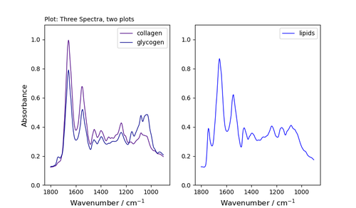
OR `fig, (ax, ax2) = plt.subplots(2,1)`
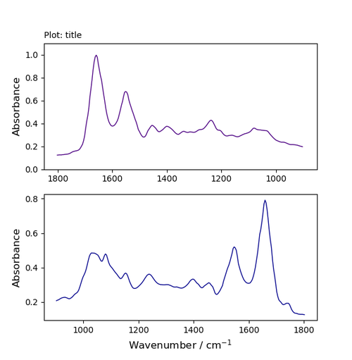
*** ** * ** ***
### [Python Script: Three Plots, Three Curves](https://midir.lightsource.ca/__attachments/a_1c243865233afcfd1f0d187494f4d4b9bdc41a1f30126f6061623ac87ed5c1b0/basic3-3.py.md?cb=3c7fffcb498dd0c0bd198a6be509f721)
Python Script: Three Plots, Three Curves
import matplotlib.pyplot as plt
import numpy as np
from Orange.data import Table
from orangecontrib.spectroscopy.data import getx
spectra = in_data.X
energy = getx(in_data) #This is specfic to getting data from Orange data files
fig, (ax, ax2,ax3) = plt.subplots(1,3)
##################################################################################################
ax.plot(energy, spectra[0], 'indigo', linewidth=1.0, label='collagen') #add label='label text' if you want to add a legend
ax2.plot(energy, spectra[1], 'darkblue', linewidth=1.0, label='glycogen') #add label='label text' if you want to add a legend
ax3.plot(energy, spectra[2], 'blue', linewidth=1.0, label='lipids') #add label='label text' if you want to add a legend
##################################################################################################
#add title
title = ax.set_title('Plot: Three Spectra, three plots',fontsize='10', loc='left') # location = centre, left, right (default is centre)
##################################################################################################
#add axis labels for left plot
ax.set_xlabel('Wavenumber / cm$^{-1}$', fontsize='12')
ax.set_ylabel('Absorbance', fontsize='12')
#add axis labels for right plot
ax2.set_xlabel('Wavenumber / cm$^{-1}$', fontsize='12')
ax3.set_xlabel('Wavenumber / cm$^{-1}$', fontsize='12')
#ax2.set_ylabel('Absorbance', fontsize='12')
#################################################################################################
#set axis limits -->can use to reverse axis for IR spectra
ax.set(xlim=(1850, 850)) #this is the wavenumber region you want to show
ax.set(ylim=(0,1.1)) #this is the absorbance range
#set axis limits for other plots
ax2.set(xlim=(1850, 850)) #this is the wavenumber region you want to show
ax2.set(ylim=(0,1.1)) #this is the absorbance range
ax3.set(xlim=(1850, 850)) #this is the wavenumber region you want to show
ax3.set(ylim=(0,1.1)) #this is the absorbance range
##################################################################################################
ax.legend() #if you added label text you need this to make a legend
ax2.legend()
ax3.legend()
##################################################################################################
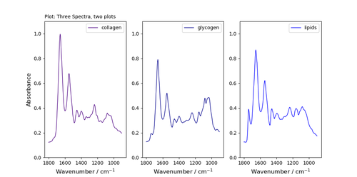
*** ** * ** ***
## [Arrows and Text](https://midir.lightsource.ca/__attachments/a_08407afc60b9248f412e56f96d539e1ef9ff83804a2f75a10d5b433f4cec739a/text_add-arrow.py.md?cb=3b4f6eeb5b219ab1d60844314126f6dd)
Python Script: Add Arrow
import matplotlib.pyplot as plt
import numpy as np
from Orange.data import Table
from orangecontrib.spectroscopy.data import getx
spectra = in_data.X
energy = getx(in_data) #This is specfic to getting data from Orange data files
fig, ax = plt.subplots()
#fig, (ax, ax2,ax3) = plt.subplots(1,3)
ax.plot(energy, spectra[0], 'indigo', linewidth=1.0, label='collagen') #add label='label text' if you want to add a legend
#ax2.plot(energy, spectra[1], 'darkblue', linewidth=1.0, label='glycogen') #add label='label text' if you want to add a legend
#ax3.plot(energy, spectra[2], 'blue', linewidth=1.0, label='lipids') #add label='label text' if you want to add a legend
#add title
title = ax.set_title('Plot: Three Spectra, three plots',fontsize='10', loc='left') # location = centre, left, right (default is centre)
#add axis labels for left plot
ax.set_xlabel('Wavenumber / cm$^{-1}$', fontsize='12')
ax.set_ylabel('Absorbance', fontsize='12')
#set axis limits -->can use to reverse axis for IR spectra
ax.set(xlim=(1850, 850)) #this is the wavenumber region you want to show
ax.set(ylim=(0,1.1)) #this is the absorbance range
#annotate
ax.annotate('key peak', xy=(1600,0.8), xytext=(1400,0.7), arrowprops=dict(facecolor='black', width=0.4, headwidth=6, shrink=0.05),)
#ax.legend() #if you added label text you need this to make a legend
plt.show()
To add an arrow, format your plot as above and then add the line:
`ax.annotate('key peak', xy=(1600,0.8), xytext=(1400,0.7), arrowprops=dict(facecolor='black', width=0.4, headwidth=6, shrink=0.05),)`
### Format the annotation
* you can add text;
* `xy` indicate the location of the arrow tip - using the plot's co-ordinates;
* `xytext` indicated the location of the text;
* choose the properties of the arrow (`facecolour`; `width`=line thickness; `headwidth` = size of head; `shrink` move the tip and base some percent away from the annotated point and text
More arrow examples can be found at
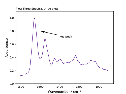
*** ** * ** ***
### Add Text
To add text to any location on your plot, include the line
`ax.text(1600,0.8, r'this is a super neat plot')`
Add text using the xy coordinates of the plot
The `r` preceding the text string is important -- it signifies that the string is a *raw* string and not to treat backslashes as python escapes.
`x-coord = 1600`
`y-coord = 0.8`
`text string = r'the text you want on the graph'`
[++Use latex++](https://matplotlib.org/tutorials/text/usetex.html) to format your text and incorporate the output directly into your figure
`ax.text(1250, .45, r'$\mu=100,\ \sigma=15$')`
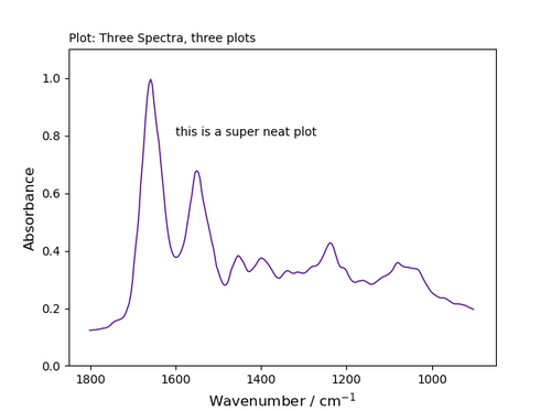
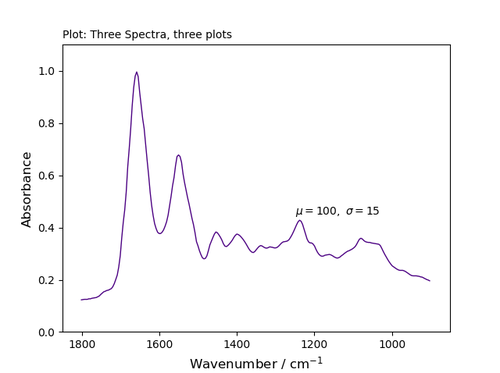
*** ** * ** ***
## Lines
| Character | Line description |
|-----------|---------------------|
| '-' | solid line style |
| '--' | dashed line style |
| '-.' | dash-dot line style |
| ':' | dotted line style |
## Markers
| Character | description |
|-----------|-----------------------|
| '.' | point marker |
| ',' | pixel marker |
| 'o' | circle marker |
| 'v' | triangle_down marker |
| '\^' | triangle_up marker |
| '\<' | triangle_left marker |
| '\>' | triangle_right marker |
| '1' | tri_down marker |
| '2' | tri_up marker |
| '3' | tri_left marker |
| '4' | tri_right marker |
| 's' | square marker |
| 'p' | pentagon marker |
| '\*' | star marker |
| 'h' | hexagon1 marker |
| 'H' | hexagon2 marker |
| '+' | plus marker |
| 'x' | x marker |
| 'D' | diamond marker |
| 'd' | thin_diamond marker |
| '\|' | vline marker |
| '_' | hline marker |
## Colours
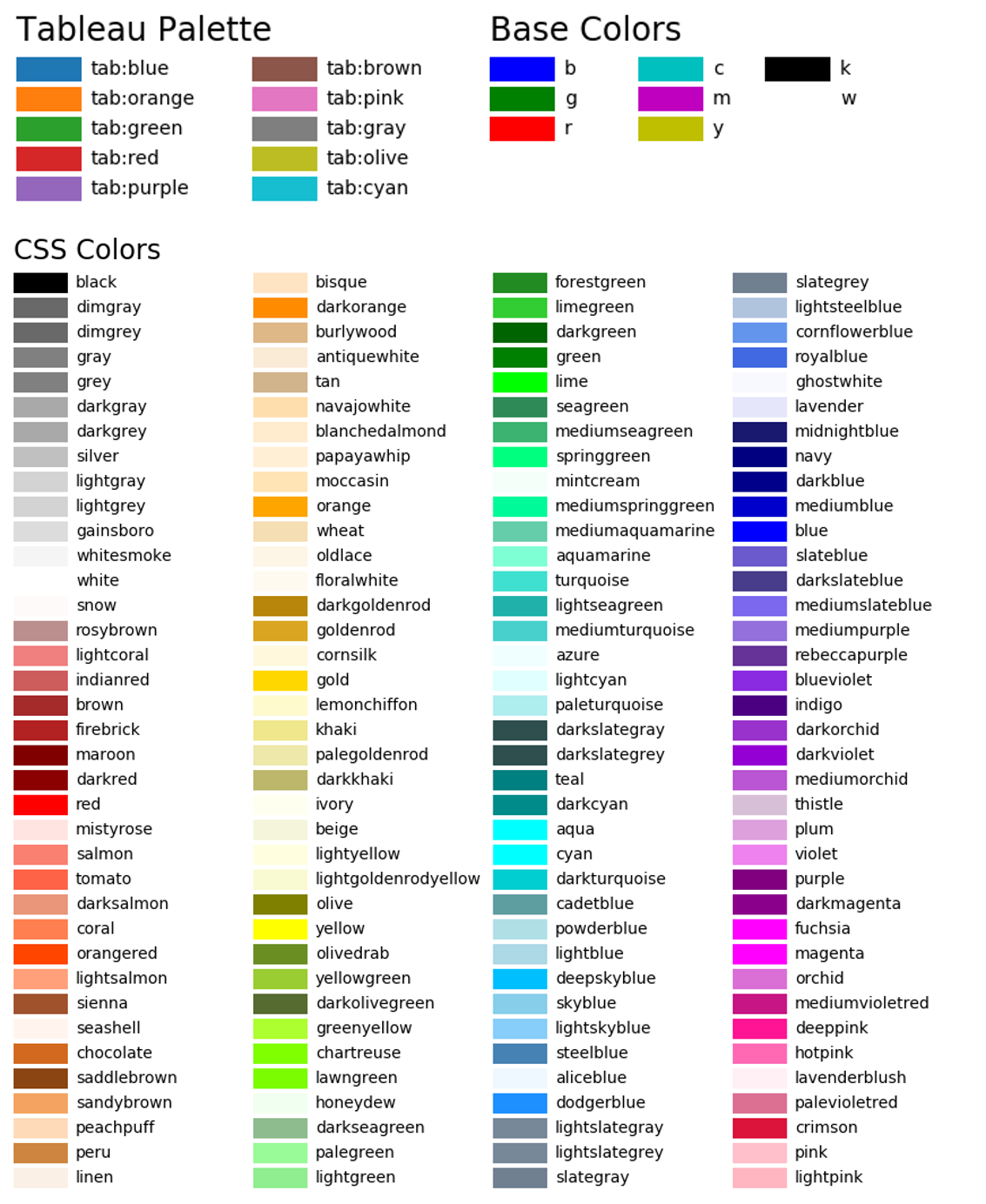
## More Options
### Want more control?
#### Layouts
Adjust the layout of your figure using `plt.tight_layout()`
#### Major and minor ticks
---
language: "en"
---
# Data Transfer
## Mid-IR Data Organization
Data collected at the Mid-IR beamline is organized by your project number. Data is further organized into two subdirectories for each project.
| **Project Folder** (example: prj12G34567) | project cycle number (12), project type (G/R/B), project number (34567) ||
| **Sub-folder names** | `raw` | `postprocessed` |
| **Sub-folder contents** | raw data collected at the endstation | modified data, user workflows, beamtime notes |
| **Sub-folder permissions: beamline computers** | read, write | read, write |
| **Sub-folder permissions: user accounts** | read-only | read, write |
|------------------------------------------------|--------------------------------------|-----------------------------------------------|
All data collected at the Mid-IR beamline is accessible on Globus and is attached to your *Canadian Light Source account and projects* . To retrieve the data, please follow this step-by-step guide. [Contact us](https://midir.lightsource.ca/mid-infrared-spectromicroscopy-mid-ir/contact.md) if you need any assistance with the data transfer.
### Access CLS Data on Globus
Globus delivers advanced file transfer and sharing capabilities to researchers no matter where their data live. [Globus](https://www.globus.org/) Connect Server makes it easy to add your lab cluster, campus research computing system, or other multiuser HPC facility to the Globus ecosystem. The CLS provides a Globus Connect Server endpoint that connects to our beamline data share, allowing users associated with scientific proposals access to their data.
### Step-by-Step Guide
#### Log in
* Access [**Globus**](http://app.globus.org/).
* Select the **Canadian Light Source** as your institution. Click **Continue**. You will receive the familiar CLS sign-in page.
* Enter your **CLS username and password**. Your first log-in will register you with Globus' systems, which is important if other users want to share with you.
* Authentication/Consent authroization may be required upon the first login. Click **Continue** to proceed.
#### File Manager \| CLS Beamline Data
* Go to the **File Manager** view and select "SEARCH"
* Search for the "**CLS Beamline Data**" Collection - this is our CLS endpoint on Globus
#### Path to Project Data
* The **CLS Beamline Data** Collection will open with your CLS username as the Path and display any project folders you are involved in.
* You will see the project you are interested in transfering data from.
#### Set-up Personal Endpoint
* In order to transfer the data to your personal computer, institution workstation or computing system you must first setup a personal endpoint. Your institution/you may already have this configured. If not:
* Navigate to [Globus Connect Personal](https://www.globus.org/globus-connect-personal)
* Install the Globus Connect Personal for your operating system
* Launch Globus Connect Personal and 'Log In' to authenticate with Globus and begin the Collection Setup process.
* Enter Collection Details for your Personal Setup and 'Save'
#### Transfer Data from CLS to Your Endpoint
* Select the files you wish to transfer to your computer
* Select the desired destination
* Click Start on CLS Beamline Data side.
* The two-pane view works well for this.
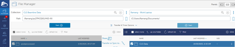
### We're here to help!
#### If you encounter any issues, please let us know. [Contact us!](https://midir.lightsource.ca/mid-infrared-spectromicroscopy-mid-ir/contact.md)
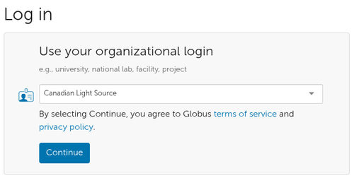
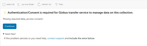
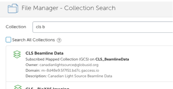
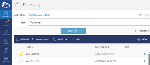
---
language: "en"
---
# Development
## Quasar is a collaborative effort of many institutions. It aims to benefit the entire scientific community through open source, democratic data analysis.
### [++Get involved!++](https://quasar.codes/contact/)
The [++public repositories++](https://github.com/quasars/) are hosted on github.
Feel free to report issues or open pull requests! Please read our suggestions on [++contributing successfully++](https://github.com/Quasars/orange-spectroscopy/blob/master/CONTRIBUTING.md).
The main repositories are:
* [++Quasar++](https://github.com/quasars/quasar) - bringing together Orange and the Spectroscopy add-on
* [++Orange Spectroscopy++](https://github.com/quasars/orange-spectroscopy) - Orange components for spectroscopy
* [++Orange++](https://github.com/biolab/orange3) - the backbone of Quasar
---
language: "en"
---
# During Beamtime
General information on what to expect can be found at [++During Your Beamtime++](https://www.lightsource.ca/users/your-cls-experiment/your-beamtime.php)
## On-site
1. Beamline staff will meet you at the beamline to help you get started with your experiment.
2. You will control all aspects of the instrument on-site
1. You have the option of using [NoMachine](https://midir.lightsource.ca/mid-infrared-spectromicroscopy-mid-ir/nomachine.md) to operate endstations remotely
3. Data will be available by method determined previous to beam time
## [++Mail-in++](https://midir.lightsource.ca/mid-infrared-spectromicroscopy-mid-ir/mail-in.md)
1. Beamline staff will run your samples using your pre-planned regions of interest (ROI)
2. Please, be available for communication
3. You will be contacted when data collection is complete
4. Data will be available by method determined previous to beam time
## [++Remote access++](https://midir.lightsource.ca/mid-infrared-spectromicroscopy-mid-ir/remote-access.md)
1. Choose method of communication with beamline staff; beamline staff will be available for communication during beam time
2. You will connect to the instrument computer using [NoMachine](https://midir.lightsource.ca/mid-infrared-spectromicroscopy-mid-ir/nomachine.md)
3. Beamline staff will setup your sample and get you oriented with the controls for the instrument
4. You will control all aspects of the instrument
5. Contact beamline staff for change samples
6. Data will be available by method determined previous to beam time
[Agilent Operation](https://midir.lightsource.ca/mid-infrared-spectromicroscopy-mid-ir/agilent-operation.md) [Bruker Operation](https://midir.lightsource.ca/mid-infrared-spectromicroscopy-mid-ir/bruker-operation.md) [Remote Access](https://midir.lightsource.ca/mid-infrared-spectromicroscopy-mid-ir/remote-access.md)
* [Mail-in](https://midir.lightsource.ca/mid-infrared-spectromicroscopy-mid-ir/mail-in.md)
[hATR Microscope Operation](https://midir.lightsource.ca/mid-infrared-spectromicroscopy-mid-ir/hatr-microscope-operation.md)
---
language: "en"
---
# FAQs
## Frequently Asked Questions
[FAQs \| General](https://midir.lightsource.ca/mid-infrared-spectromicroscopy-mid-ir/faqs.md#General)
[FAQs \| Proposals](https://midir.lightsource.ca/mid-infrared-spectromicroscopy-mid-ir/faqs.md#Proposals)
[FAQs \| Sample Preparation](https://midir.lightsource.ca/mid-infrared-spectromicroscopy-mid-ir/faqs.md#Sample-Preparation)
[FAQs \| Mail in/Remote Access](https://midir.lightsource.ca/mid-infrared-spectromicroscopy-mid-ir/faqs.md#Mail-in/Remote-Access)
[FAQs \| Data](https://midir.lightsource.ca/mid-infrared-spectromicroscopy-mid-ir/faqs.md#Data)
## General
> My laboratory has an infrared spectrometer and/or infrared microscope. What are the advantages of collecting data at Mid-IR Beamline?
Compared to a standard lab spectrometer that collects a spectrum averaged over a large area, the imaging microscope at Mid-IR collects a map of infrared spectra. Visit the [++About Us++](https://midir.lightsource.ca/mid-infrared-spectromicroscopy-mid-ir/about-us.md) page to find out specifics about the instruments at Mid-IR.
Curious about other advantages? We offer:
**High spatial resolution**
**Excellent signal to noise**
**Large field of view**
**Fast data collection**
**Infrared expertise in data collection and analysis**
> Our group has never done infrared spectromicroscopy before - is it appropriate to use the CLS for our research?
Yes! It is a goal is to attract new users to the CLS and expand the user community. Please [++contact us++](https://midir.lightsource.ca/mid-infrared-spectromicroscopy-mid-ir/contact.md) to discuss!
> What wavelengths does Mid-IR beamline cover?
The Mid-IR beamline covers 560 - 6000 cm ⁻¹ ( 70 - 744 meV )
> How long will it take to measure an IR map/image of my sample?
The length of time required to collect an infrared measurement, image or mosaic will really depend on the *size of the region of interest* and the *collection parameters* (resolution, # of scans, binning, etc). Ask staff for help with estimating your measurement time.
> I want to run the same sample at multiple beamlines, can I do that?
You sure can! We collaborate with many of the x-ray beamlines. Please [++contact us++](https://midir.lightsource.ca/mid-infrared-spectromicroscopy-mid-ir/contact.md) to discuss specific experiments.
*** ** * ** ***
## Proposals
> How do I answer "need for IR synchrotron radiation?"
When using mid-infrared radiation, the only advantage provided by a synchrotron source is given by its high brightness. Brightness is critical when performing mapping and spectromicroscopy experiments requiring diffraction limited spatial resolution and high signal/noise ratio. Your proposal must explain clearly why your scientific problem requires such performance.
> How do I make efficient use of beam time?
Create a detailed experimental plan. List samples in order of priority, plan number and locations of region of interest, understand data collected from complimentary techniques.
If this is your first time doing synchrotron or infrared spectromicroscopy measurements discuss with collaborators or colleagues and beamline staff about your plan and whether or not is it reasonable.
*** ** * ** ***
## Sample Preparation
> Do I need to microtome my samples?
The CLS has a microtome on site that can you can use. Please indicate in your GUP proposal you would like to use it and contact beamline staff to discuss details.
> Can I wash my IR windows?
Yes, except for barium fluoride windows. IR windows can be reused a few times before they become scratched. Wash with water or suitable alcohol-based solvents and dry gently with lens tissue.
> How thick should my sample be?
Sample thickness generally depends on the measurement mode (transmission, ATR, etc). For transmission 5 - 10 µm sample thickness if possible; in ATR and reflectance the IR light does not transmit through the sample, and other parameters such as roughness are more critical. The [++sample++](https://midir.lightsource.ca/mid-infrared-spectromicroscopy-mid-ir/samples.md) section has more information and please [++contact us++](https://midir.lightsource.ca/mid-infrared-spectromicroscopy-mid-ir/contact.md) about specific sample requirements.
*** ** * ** ***
## Mail-in/Remote Access
> My beamtime is coming up, how do I choose between mail-in and remote access?
Currently, we recommend on-site access whenever possible. Please [++contact us++](https://midir.lightsource.ca/mid-infrared-spectromicroscopy-mid-ir/contact.md) and we will discuss the specifics of your experiment.
> My samples need to be refrigerated or frozen, is this possible?
No problem! Please indicate on your shipping forms and inform us ahead of time. There are options of
* Refrigerator (5 **°**C)
* Freezer (-20 **°**C)
* Extra cold freezer (-80 **°**C)
Check out [++our facilities++](https://www.lightsource.ca/facilities/laboratories-equipment.php) for more information about the labs and equipment we have on-site.
*** ** * ** ***
## Data
> How do I analyze the infrared data collected?
The CLS Mid-IR Beamline uses, [develops](https://midir.lightsource.ca/mid-infrared-spectromicroscopy-mid-ir/development.md) and supports, [++Quasar++](https://midir.lightsource.ca/mid-infrared-spectromicroscopy-mid-ir/software.md) -- an open-source collection of spectroscopic data analysis toolboxes extending the [++Orange machine learning++](https://www.tandfonline.com/doi/full/10.1080/08940886.2017.1338424) and data visualization suite. ([++Orange, 2013++](https://jmlr.org/papers/volume14/demsar13a/demsar13a.pdf)) The suite of powerful tools has visual data analysis workflows to support novice and expert users alike in creating dynamic and reproducible data pipelines which integrate spectral processing, hyperspectral maps and multivariate analysis.
More information can be found [++here++](https://midir.lightsource.ca/mid-infrared-spectromicroscopy-mid-ir/software.md). To cite: [++Quasar - Citation++](https://quasar.codes/citation/).
> I have my own data analysis routines written in Python, can I use them in Quasar?
Yes! Please check out the [++custom code++](https://midir.lightsource.ca/mid-infrared-spectromicroscopy-mid-ir/custom-code.md) section for information on how to implement your routines.
---
language: "en"
---
# Focal Plane Array
Focal Plane Array (FPA) detectors allow for the simultaneous acquisition of an n by n number of spatially resolved spectra, and each pixel provides an infrared spectrum. Thousands of spectra are simultaneously acquire within minutes. Each n by n image is called a tile.
Infrared images of large regions of interest can be performed by collecting many tiles of data and automatically stitching them together to create a composite mosaic.
The [++**Agilent**++](https://midir.lightsource.ca/mid-infrared-spectromicroscopy-mid-ir/agilent.md) is equipped with a 128 x 128 pixel FPA and the [++**Bruker**++](https://midir.lightsource.ca/mid-infrared-spectromicroscopy-mid-ir/bruker.md) with a 64 x 64 pixel FPA
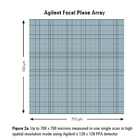
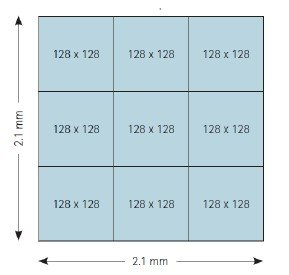
FPA detectors are liquid Nitrogen-cooled. For the Agilent endstation, we use an automatic Liquid Nitrogen delivery system to refill the detector at timed intervals. For the Bruker, refills must be done by users and/or staff, requiring on-site activity. Beamline staff will instruct you on proper cooling schedules and best practices at the beginning of your beamtime when using these detectors.
---
language: "en"
---
# Getting Started
## Where to begin
### So you find yourself with beautiful data from your beam time at Mid-IR, now what?
Quasar is an excellent program for viewing and analyzing your data whether this is your first time with infrared data or you're a seasoned pro.
Quasar offers spectral processing routines including baseline subtraction, normalization, FFT, EMSC, peak analysis, differentiation and smoothing which can be quickly and easily applied to a dataset.
*** ** * ** ***
## Installing Quasar (preferred)
The preferred installation method is to use the pre-built Quasar installers.
Quasar Download \& Installation Guide
1. Download Quasar from:
2. Run the installer \& follow prompts
1. The installer includes the `opusFC` and `orange-spectroscopy` packages by default!
3. On to Science!!
## Installing from Orange (alternative)
If you are already using Orange or prefer to use it instead of Quasar
Orange Download \& Installation Guide
1. Download Orange from:
2. Run the installer, which will also install the miniconda python distribution.
3. Install `orange-spectroscopy` by going to "Options" "Add-ons..." and choosing "Orange-Spectroscopy".
4. If you want to read OPUS files, you must manually install the opusFC package:
1. Go to Options / Add-ons
2. Click the "Add more..." button
3. Enter "opusFC" into the text box
4. Check the box next to "opusFC" and click OK to install
*** ** * ** ***
## Additional Resources
Reach out for [++help++](https://midir.lightsource.ca/mid-infrared-spectromicroscopy-mid-ir/contact.md)
[++Getting started guide (Orange)++](https://orange.biolab.si/getting-started/)
Watch some [++tutorials++](https://midir.lightsource.ca/mid-infrared-spectromicroscopy-mid-ir/tutorials.md)
Dive into some example [++workflows++](https://midir.lightsource.ca/mid-infrared-spectromicroscopy-mid-ir/workflows.md)
Learn some of our [++tips and tricks++](https://midir.lightsource.ca/mid-infrared-spectromicroscopy-mid-ir/tips-and-tricks.md)
---
language: "en"
---
# hATR Microscope
## Bruker Endstation: Custom Horizontal ATR Microscope
### 🟡 Special Request using the Bruker Endstation
The horizontal ATR microscope is designed to provide convenient horizontal sample orientation for challenging samples measured using ATR combined with off-axis parabolic (OAP) focusing optics for optimal illumination of a variety of Internal Reflection Elements (IREs). This endstation can be coupled to both the synchrotron and globar light sources with ample opportunity for customization of both sampling environment and measurement geometry. [Contact our Beamline Responsible](mailto:scott.rosendahl@lightsource.ca), Scott Rosendahl, to inquire how this microscope may be configured for your experiment.
### Measurement Configurations
MCT single-point spectroscopy, 150 µm pinhole at conjugate focus
FPA imaging, 3x magnification, FOV is 850x850 µm with 13.3x13.3 µm pixels, expanded by cos(θ~AOI~)^-1^ along the optical axis, composite stitching available.
### Further Reading
See the experimental section of this paper for more details: Morhart, T. A.; Read, S.; Wells, G.; Jacobs, M.; Rosendahl, S. M.; Achenbach, S.; Burgess, I. J. Attenuated Total Reflection Fourier Transform Infrared (ATR FT-IR) Spectromicroscopy Using Synchrotron Radiation and Micromachined Silicon Wafers for Microfluidic Applications. *Appl Spectrosc* **2018** . [++doi:10.1177/0003702818785640++](https://doi.org/10.1177/0003702818785640).
### Techniques
**Synchrotron ATR-FTIR Spectroscopy**
**ATR Infrared Chemical Imaging**
### Detectors
**Single-element 250 µm Kolmar MCT**
[++**Focal Plane Array**++](https://midir.lightsource.ca/mid-infrared-spectromicroscopy-mid-ir/focal-plane-array.md)**(64 x 64)**
---
language: "en"
---
# hATR Microscope | ATR Imaging
## Return to [++hATR Microscope Operation++](https://midir.lightsource.ca/mid-infrared-spectromicroscopy-mid-ir/hatr-microscope-operation.md) User Guide
## Using the FPA Detector
This detector needs to only be operated at appropriate temperatures -- you must refill the detector with liquid nitrogen at least every **8 hours** or risk damaging it. Your beamline staff will show you the liquid nitrogen refill procedure. The FPA detector can never be on when it is warm -- if you plan to leave the beamline unattended for a longer duration you must **turn off the detector before leaving**. When turning off/on the detector, make sure OPUS software is closed. Once the detector is off/on the software can be restarted.
## Planning Your Measurements
Raw images will undergo post-collection processing to transform *pixel* indexes into meaningful sample measurements. The microscope magnification and angle of incidence along with the internal reflective element (IRE) refractive index and geometry and the detector pixel sizes will need to be accounted for in the routine.
## Data Collection Workflow
1. **Data Collection:** Raw images will be collected by the detector in terms of *pixel indeces* . You can glance at these raw images, but they will contain little spatial information with respect to your sample, and are *rotated* by the angle of incidence of the microscope. They are also compressed in the optical axis. Images will be planned at specific sample locations by the Wizard application. The image locations are determined by a calculation performed by the Wizard with cropping height, cropping width and effective angle of incidence calculated from user inputs. The software saves the stage location in the file name of each collected image. The software will also save the measurement details such as effective AOI, microscope set angle, cropping width, cropping height.
2. **Image Post-Processing:**
1. **Image correction:** rotation, elongation in reflection axis and conversion from pixel index to sample coordinates. The effective angle of incidence, microscope set angle, cropping width and cropping height are used in this stage. Orange scripts will read this information from collected files and input into the image correction algorithms.
2. **Image cropping:** Pixels are selected from the most in-focus, central region of each image. The crop x/y inputs are used at this stage. This defines the height/width of each 'tile' from the respective images.
3. **Image stitching:** Cropped images are combined to create a large composite image of the sample.
## Data Collection and OPUS Set-up
### HTTP OPUS
A special version of OPUS exists specifically to navigate commands from the hATR Wizard. If you are planning to use the Wizard for measurements, make sure you open the OPUS in the Temp folder on the Bruker computer's Desktop.
### XPM File
The HATR-FPA.xpm file should be loaded into the Advanced Data Collection tab -\> if you wish to change the file save location make sure to *overwrite* the HATR-FPA.xpm file in the HTTP/temp folder. This file will be used by the Wizard to measure. The number of scans and the sample namecan be overwritten by the Wizard program, but you might want to adjust these to suit your desired experiment and set defaults.
### Troubleshooting OPUS
What to do if OPUS crashes? Shut down the software and try to reopen the OPUS in the Temp folder. If 'another instance of OPUS is already running' shows up try to shutdown the computer and restart it with the power button on the computer. Refreshing the start-up may solve the issue. If not, contact your beamline staff!
What to do if you see red error symbols in the FPA Image Acquisition window? Try opening the single file method editor and loading the `HATR-FPA.xpm` file. Then move back to the FPA Image Measurement and reload the correct .xpm file. If nothing changes, contact your beamline staff -- it's possible the spectrometer and/or OPUS may need to be restarted.
Make sure the last collected background position has the same resolution as your measurement grid settings if the ABS data block is selected in your XPM file.
## Wizard Set-up
### Regions of Interest
Add measurement points of interest. 'Add ROI Point' button will add a point at the current stage positon to the planned region. Navigate around your sample to add further region of interest points. The software will surround all points added in the region of interest with sample tiles from inputted crop x/crop y settings. Points may also be added by clicking on the ROI Bokeh plot.
### Determining Pixel Cropping
Crop X (px) and Crop Y (px) are set in the hATR Wizard software. Adjust the pixel cropping to compromise between measurement time and image quality. Starting values should be set to the Wizard's default parameters and can be changed by the user. Use the Wizard/image area as a guide for how long the measurement will likely take and the expected image area. Adjusting these values will adjust the *width* and *height* of each FPA measurement/tile.
### Time Estimator
Just underneath the planned measurement locations there is an 'Estimate' Accordian where you can see the estimated timing for the current ROI plan with the selected Resolution and Scans set in the drop-down menu. Once you have decided these paramters, remember to update the [OPUS Settings](https://canadianlightsource.atlassian.net/wiki/spaces/MIDIR/pages/415007370/hATR+Microscope+Set-up+and+User+Guide#Wizard-OPUS-Settings) to reflect these changes.
### Wizard OPUS Settings
Scrolling down to the bottom of the left pane panel, you will see inputs underneath the 'OPUS Settings' title. Update the **Sample name** , **Resolution** , and number of **Scans** to reflect your desired experiment. These will be used by the software to *overwrite* the default OPUS settings.
### Saving and Restoring Wizard Session
At the very bottom of the panel, you will see the Save/Restore buttons:
Save → Saves the current Wizard session to the Cache.
Restore → Upon reloading/opening the hATR wizard program, hit the 'Restore' button to restore the last saved session.
## Planning Measurement Positions
### Add ROI Points
1. Add ROI Point Button: This green button (outlined in red in the above image) allows the user to Add a new ROI point to the measurement plan. It will append the *current stage location*as an ROI point.
2. Edit ROI Points Button on Bokeh Plot: The three dots surrounding an arrow button is located above the Bokeh plot 'Positions' Tab. This Bokeh plot provides a visual showing the ROI points (red) and the measurement locations planned by the software (red box outlines). The software takes your ROI points and surrounds them with measurement 'tiles' in a grid formation - taking into account your set pixel cropping settings and the effective angle of incidence set above. In the example, the software is planning a 10-tile measurement plan (2-tiles in X and 5 tiles in Y). Each box represents one measurement location, one *file* to be collected by OPUS.
### Add New Region
Use the blue ➕ button to add a new region → this will append the new region to the selection drop-down menu and you will have a fresh view on your Bokeh plot. This can help with measurement planning when multiple measurement regions are desired for the same sample.
### Measure Grid
The blue button, 'Measure Grid' will start the OPUS acquisition, moving the stage to each of the XY locations in the planned array and acquiring an FPA image at each location.
### Typical workflows
1. One background, multiple sample measurements at different locations. Perhaps your background is of the bare crystal and the sample on top of the crystal at different locations. It is recommended that you stay close to the center X position for these (+/-1250um in X is reasonable). In this case, the background can be collected first at the background location. Once the sample is in place you can collect a grid/series of measurements of your sample.
2. A background measurement and sample measurement at each measurement location. Perhaps you measure a grid with the bare crystal and obtain a matrix of data. Then with the sample in place you measure the same exact area again. In this case, the absorbance data will have to be calculated after data collection. This type of dataset could also be used to compare two measurement conditions to eachother i.e. a battery before/after discharging/charging. The same locations on the crystal with different temperature conditions applied to them, you name it!
### Save Stage Positions
This feature allows one to save XYZ stage locations into the Wizard. In some cases, fine-focusing may be required for your sample and you may want to reference the XYZ position for later use. If you find a focal point you want to reference later this would be a good place to save the point. As of now, there is no naming system -- this would be a nice update to this feature. Save stage positions allow for measurement planning, crystal orientation
## Quick Set-up
1. Input IRE and microscope information into the hATR Wizard
2. Adjust Crop X and Crop Y if desired
3. Add ROI points to define image area of sample
4. Change the name of new regions of interest for easier data processing and sorting
5. Measure Grid
6. Process Data in Quasar
### OPUS Data Blocks
`ABS` -- absorbance of the sample measurement (`-log[SSC/RSC]`) -- it's only beneficial to save absorbance data if you only intend to have one background position collected at the beginning of the dataset.
`SSC` -- single channel of the sample measurement -- it's best to always save this data in case you need to reprocess afterwards
`RSC` -- single channel of the reference measurement
Tip: If you are saving absorbance (`ABS`) data blocks you must collect a background measurement in OPUS before commanding the Wizard to measure an area.
Tip: Collect a background measurement at the achieved focus position. Then deposit a water droplet onto the FAC surface and collect a small grid to see where the water is and to *resolve the droplet boundary conditions.*This provides a guide in selecting an appropriate cropping settings.
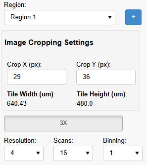
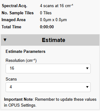
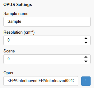
Tip: If you are planning to save the Absorbance data block you **must** make sure the background measurement was obtained with the *same resolution* as you set for your sample grid.
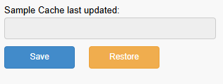
Tip: At regular checkpoints during your experiment, remember to hit 'Save' so you can re-load your session if the program crashes/closes for some reason.
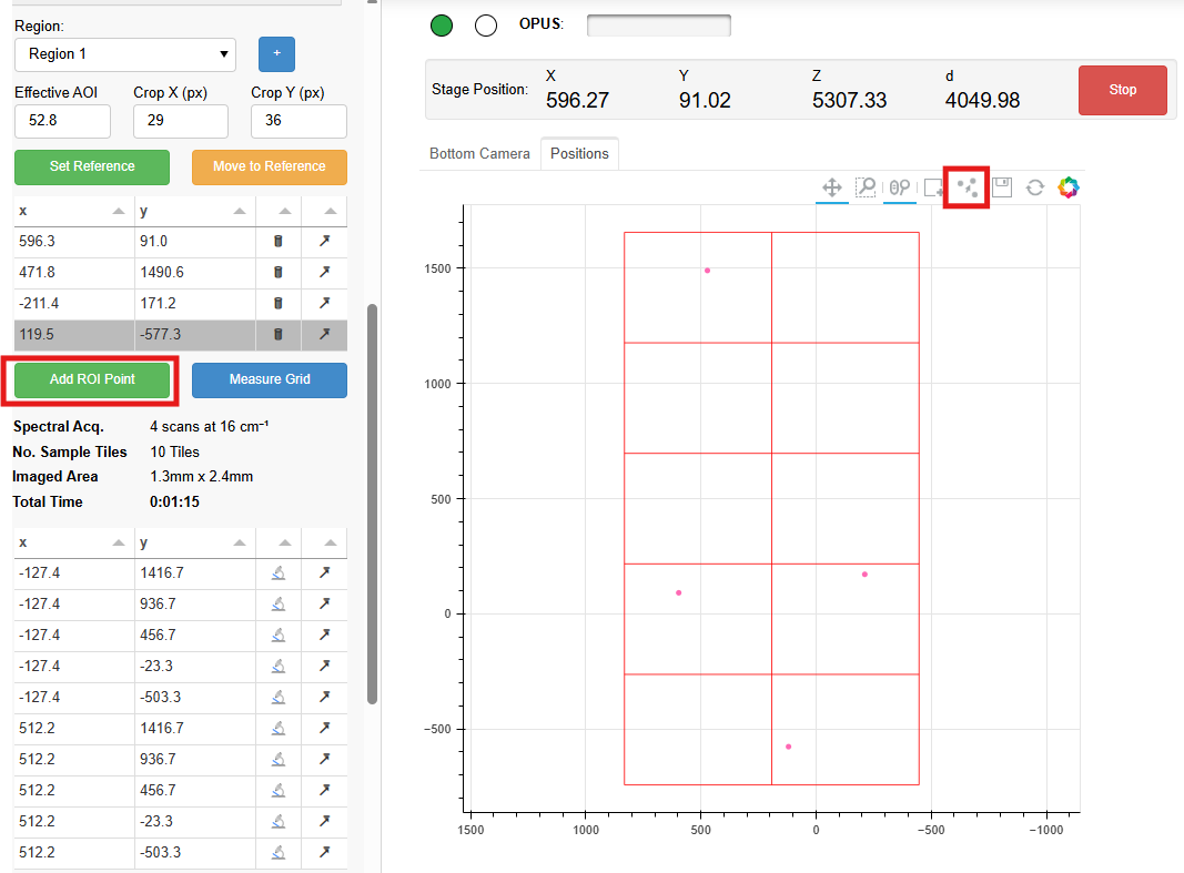
Tip: Make sure your OPUS XPM file and Wizard OPUS Settings are set before committing to the measurement. Contact your beamline staff if you run into an issue with this.
---
language: "en"
---
# hATR Microscope | MacroIRE Setup
## Return to [++hATR Microscope Operation++](https://midir.lightsource.ca/mid-infrared-spectromicroscopy-mid-ir/hatr-microscope-operation.md) User Guide
## Internal Reflection for Macro IREs
The stage is aligned for Macro IREs, such as FACs, such that external reflection is achieved at the desired IRE/Sample interface height. Depending upon the geometry and material of the IRE, light will take a different path through the crystal than compared to through air (i.e. external reflection). We must translate the crystal such that we achieve total internal reflection at the absolute **center** of the principal reflection plane of the FAC.
Total internal reflection occurs when a high refractive-index material is in place, like a face-angle crystal or similar, and the light approaches the Sample/FAC interface at an angle equal to or above the critical angle.
We can translate the stage such that total internal reflection will be achieved through your IR once the following information about your sample is known: **material, size, face angle, diameter and height.** To move from external (through-air) to internal (through-high-refractive index media) one must move the sample *away* *from the source* in both X and Z by the calculated amount. This means we *increase* X and Z by the amounts. Additional translations are required for the receiving optics attached to a rail with position D. One must also increase D by the amount calculated -- D will be moved *away from the source*.
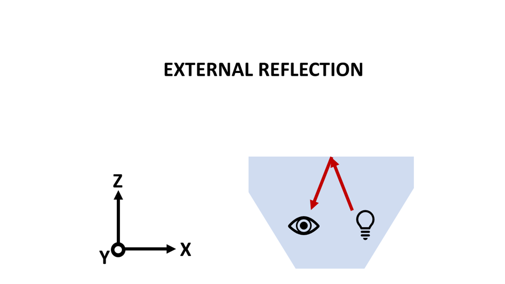
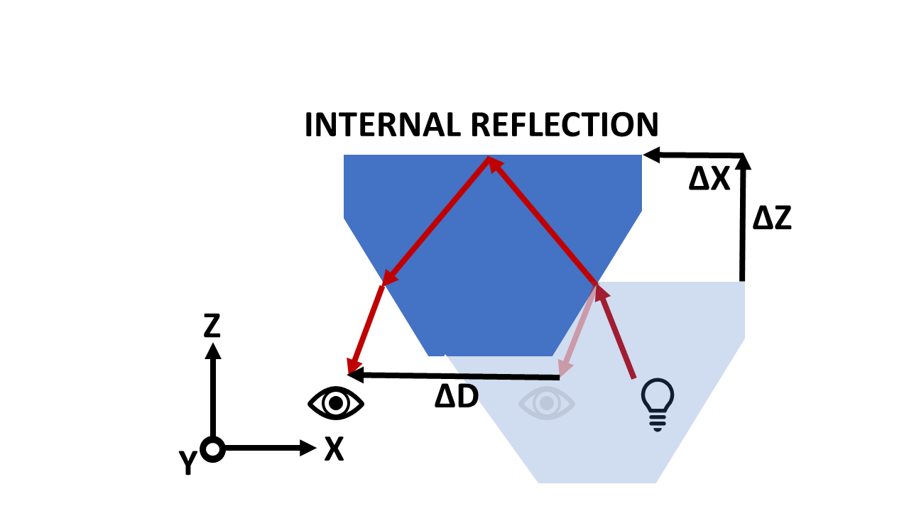
## Procedure
Staff will help with this initial set-up.
1. Determine external reflection focus at sample/IRE plane -- use mirror on IRE/sample plane. Mirror may be used in the center of the FAC holder. Lower/raise stage z and adjust d to find the focus point, adjust y axis to be at IRE center-point -- test with mirror bisecting FAC holder.
Set Current Stage Position XYZ External Reflection
Set Current Rail Position D External Reflection
2. Place FAC in holder, ensure proper alignment of FAC in holder -- IRE seated into holder properly, IRE face angle perpendicular to beam path, holder fastened to stage adapter plate, stage adapter plate properly seated.
3. Enter the details for your sample and IRE including:
Microscope set angle (35° unless adjusted)
IRE face angle (45° / 60° / other)
IRE material real refractive index
Sample real refractive index (keep as 1)
Make sure the above details are correct in the wizard before proceeding with alignment. The wizard will calculate the effective angle of incidence and the crystal translations to align your system in internal reflection. These values are in micrometers.
4. Translate crystal XYZ and collection optics D using the two yellow Move buttons -- note that these translations will be unique to each FAC material, FAC geometry and microscope set angle.
5. Verify focus
6. Proceed to measurements
### Measurement Area
The center of the FAC (in x and y) is the ideal location for measurement. Some experiments may desire a range of measurement positions such as linescans, point-by-point mapping and imaging of larger regions of samples. The sample stage may be translated in Y for at least 1 cm total range of motion (5 mm on either side of due center). This cut-off is realized due to the geometry of the sample holders. The sample stage may be translated in X away from/toward the source. This results in distortion of the beam through the FAC -- the entering beam and exiting beam travel different distances through the high-refractive index medium, causing distortion. Deviations from center will exhibit loss of focus/spatial distortion. It is recommended to stay within 3 mm of center -- 1.5 mm in either direction. Therefore, the measurement area is confined to the central 10 mm x 3 mm (Y and X).
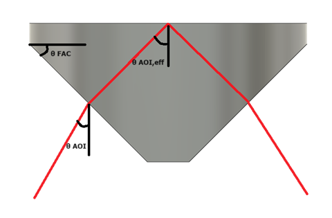
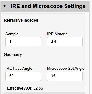
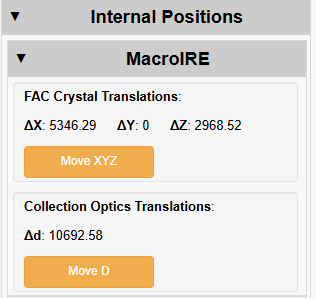
---
language: "en"
---
# hATR Microscope | MicroIRE Setup
## Return to [++hATR Microscope Operation++](https://midir.lightsource.ca/mid-infrared-spectromicroscopy-mid-ir/hatr-microscope-operation.md) User Guide
## Internal Reflection for Micro-IREs
The stage is aligned for micro-IREs to be externally reflecting off the *bottom terraces* of the microgrooves. We have developed an optical/geometrical analysis and calculator in the hATR Wizard to guide internal reflection positions for microIREs. The XYZ and D translations are unique to each IRE material, microgroove geometry and microscope set angle. Input the following information about the micro-IRE into the hATR Wizard: material, size - including thickness, groove depth, facet positioning. The IRE is translated *towards the source* in Z (decreasing stage Z) and *away from the source* in X (increasing stage X). The D-rail will also be moved *away from the source*. Both translations may be completed with buttons in the Wizard.
## Procedure
Staff will guide you through the following steps to set up your system for measurements.
1. Determine external reflection reference position. For microIREs, this is the center point of a terrace (bottom of groove) of the IRE.
Find the appropriate z/d positions for external reflection on IRE base -- use mirror / flip IRE / use sample area away from terrace features for focus
2. Place crystal in holder, ensure proper alignment -- IRE seated into holder, IRE face angle perpendicular to beam path, holder fastened to stage adapter plate, stage adapter plate properly seated to stage.
Find the x position where the beam is centered on the IRE terrace -- use visible laser / FPA / MCT / QCL signals as feedback
1. Set Current Stage Position to XYZ External Reflection
2. Set Current Rail Position D External Reflection
3. Enter the details for your sample into the hATR Wizard:
1. IRE and Microscope Settings:
1. IRE material real refractive index
Sample keep at 1
IRE face angle (custom°)
Microscope set angle (35°)
2. MicroIRE Geometry Settings (in mms):
IRE height (thickness)
IRE gap (width between edge of terraces)
IRE terrace (width of base in between grooves)
IRE ledge (distance from IRE/sample surface to groove apex)
3. ⚠️ Verify the delta positions are suitable for your system. The values are calculated in micrometers. The Wizard will calculate the required crystal and collection optics translations to align your sample into internal reflection.
4. Translate the crystal in XYZ and the collection optics in D using the Move buttons.
5. Verify proper focus and proceed to measurements.
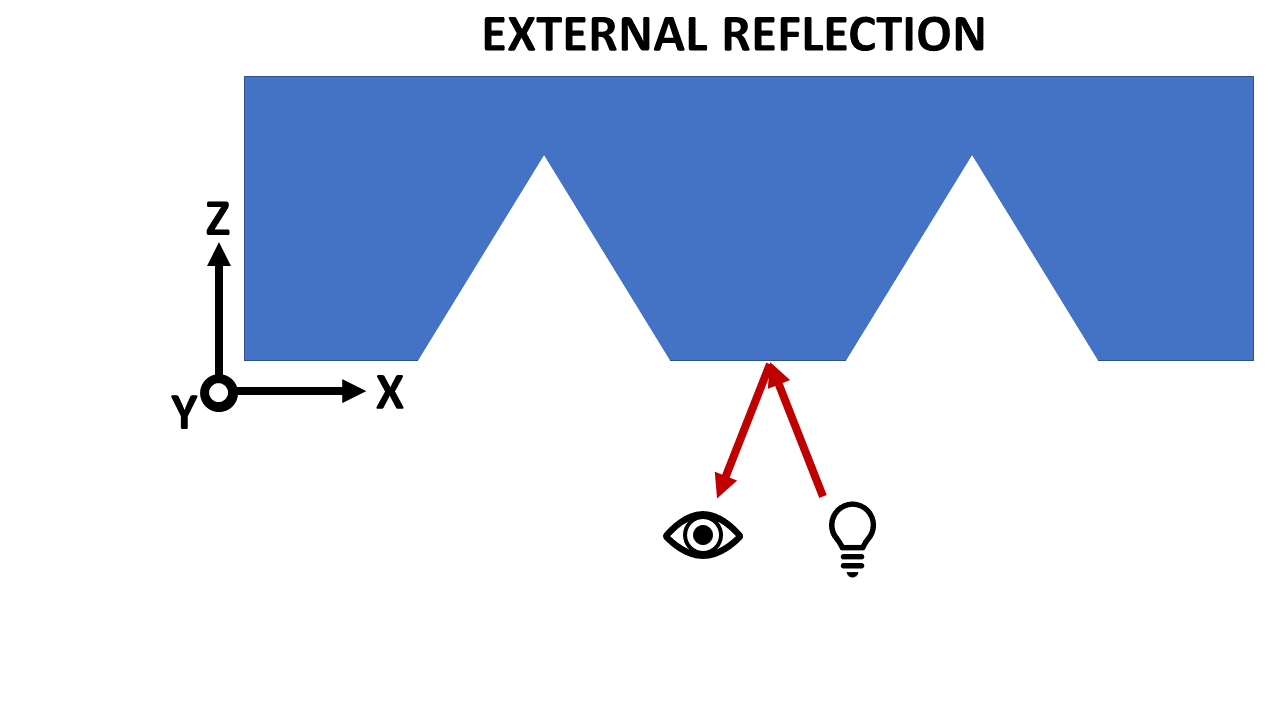
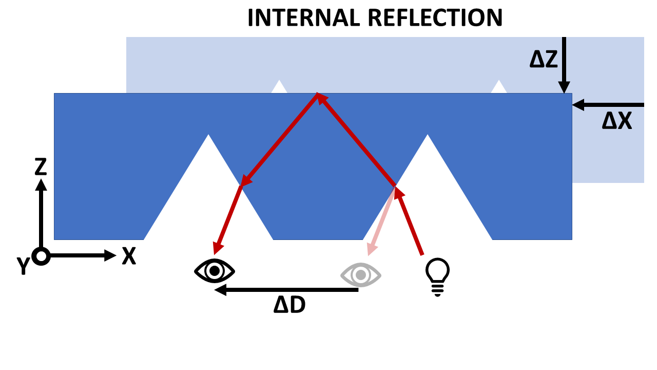
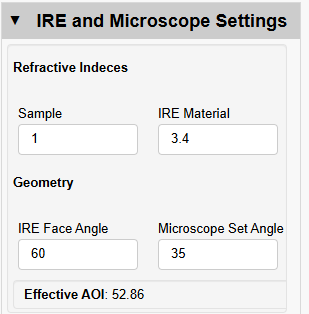
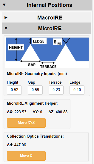
---
language: "en"
---
# hATR Microscope Operation
## hATR Microscope User Guide
[**About the hATR Microscope**](https://midir.lightsource.ca/mid-infrared-spectromicroscopy-mid-ir/hatr-microscope.md)
**ATR Imaging**
**Synchrotron FTIR**
**More Information**
### Instrument Orientation
#### Detectors and Liquid Nitrogen
Both the single point mapping (MCT) and imaging (FPA) detectors are cryogenically cooled for operation with liquid nitrogen. A single detector fill will last at least **12 hours** for the MCT and up to a maximum of **8 hours** for the FPA.
* It is very important **not to allow the FPA detector to warm up** while is it powered on.
* The MCT should be allowed to thermalize for **20-30 minutes** after filling.
The detector will be initially filled with liquid nitrogen for you by beamline staff, however you will need to plan your experiments to accommodate refilling the detector.
Appropriate **PPE** is available at the beamline and must be worn whenever handling liquid nitrogen. If you are unsure how to fill the detector or have never done it, ask a member of the beamline staff to demonstrate. You should also have completed the **cryogenics safety training** module.
#### Spectrometer and Microscope
The instrument consists of a spectrometer (Vertex 70v, on left), the transfer optics (plexiglas box in center), and the custom horizontal ATR microscope (hATR). Synchrotron radiation enters the spectrometer from the rear out of the active optics chicane.
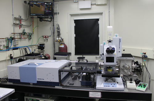
#### Sample Stage Controls
The hATR microscope sample stage can be controlled by a customized 'hATR Wizard'. The sample position can also be adjusted with the XYZ rotating knobs. The D-rail, which hosts the collection mirrors, can also be controlled with the hATR wizard.
#### Microscope Coordinate System
Z → vertical direction, raising/lowering the sample away from  or towards  the incoming light source.
X → direction parallel to beam path.
Y → direction perpendicular to beam path. Changes in this direction will not adjust sample 'focus'
D → collection optics (receival OAP and corresponding flat) are mounted on our d-rail. The rail can be moved toward  or away from  the incoming light source.
## Navigating Your Sample
Note on sample stage movement: the beam position is *fixed* and we translate the sample. Below is a descriptive view of translating a mirror with the stage.
### X Direction: Moving Sample Parallel to Beam Path

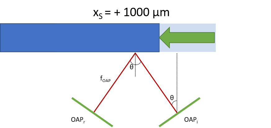
Translating the sample *away from source* .
Sample side *nearest beam* is illuminated.
🟰
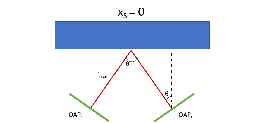
Sample center illuminated by beam.

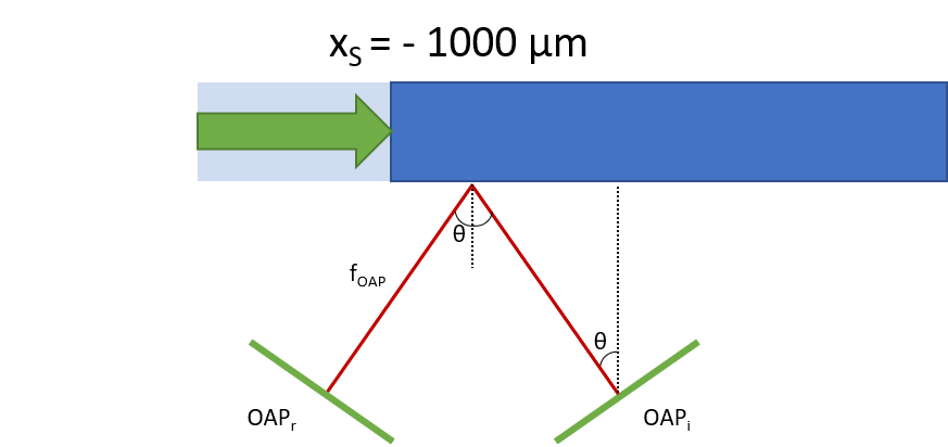
Translating the sample *towards the source* .
Sample side *furthest from beam* is illuminated.
### Z Direction: Moving Sample Vertically

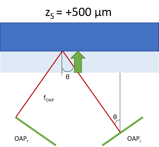
Raising the sample *away from source*.
🟰
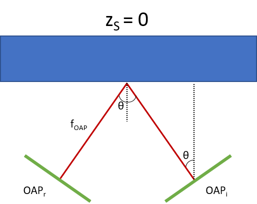
Mirror center illuminated in external reflection

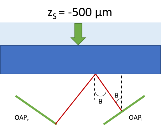
Lowering the sample *towards the source.*

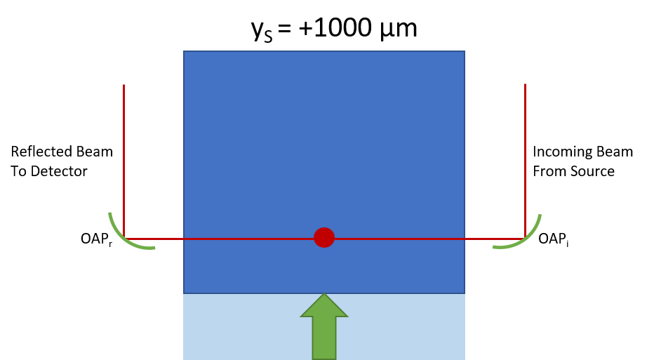
🟰
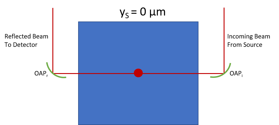

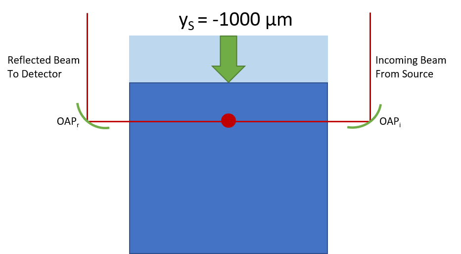
## Experimental Setup
These steps should be completed for you by beamline staff, however, you should confirm the detector cooling before starting your work.
1. Fill the detector(s) you will use for your experiments. If you're using the FPA, turn it ON after it is cooled to liquid nitrogen temp.
2. Confirm the experimental geometry with staff.
3. Start OPUS using the icon in the temp folder on the Bruker desktop.
4. Configure Transfer Optics for experiment.
5. Run the hATR wizard using the Bruker desktop shortcut.
### Load Sample onto Sample Stage
### Experimental Workflows
The system is now ready to be set-up for your desired experimental workflow.
* [hATR Microscope \| MacroIRE Setup](https://midir.lightsource.ca/mid-infrared-spectromicroscopy-mid-ir/hatr-microscope-macroire-setup.md)
* [hATR Microscope \| MicroIRE Setup](https://midir.lightsource.ca/mid-infrared-spectromicroscopy-mid-ir/hatr-microscope-microire-setup.md)
* [hATR Microscope \| ATR Imaging](https://midir.lightsource.ca/mid-infrared-spectromicroscopy-mid-ir/hatr-microscope-atr-imaging.md)
---
language: "en"
---
# Mid Infrared Spectromicroscopy (Mid-IR)
The Mid Infrared Spectromicroscopy beamline provides a state-of-the-art Fourier Transform IR spectrometer and microscope to supply diffraction-limited spatial resolution to an ever-widening range of experimental configurations. Research and development will explore new experiments and re-examine existing techniques by applying the advantages of high brightness infrared synchrotron light. Learn more [About Us](https://midir.lightsource.ca/mid-infrared-spectromicroscopy-mid-ir/about-us.md)!
## **Beamline Announcements**
The [**Call for Proposals**](https://www.lightsource.ca/users/user-information.php) is OPEN for Cycle 43 beamtime in Jan-June of 2027! The call will be CLOSING on Aug 19, 2026. [Contact](https://midir.lightsource.ca/mid-infrared-spectromicroscopy-mid-ir/contact.md) beamline staff to discuss projects and plan your [proposal submission](https://midir.lightsource.ca/mid-infrared-spectromicroscopy-mid-ir/preparing-a-proposal.md)!
Check out out our [News](https://midir.lightsource.ca/mid-infrared-spectromicroscopy-mid-ir/news.md) page for the latest [beamline news](https://midir.lightsource.ca/mid-infrared-spectromicroscopy-mid-ir/beamline-news.md) and [research highlights](https://midir.lightsource.ca/mid-infrared-spectromicroscopy-mid-ir/research-highlights.md). Normal operations in [**Cycle 42**](https://www.lightsource.ca/users/operations-schedule.php) are underway on the [Agilent](https://midir.lightsource.ca/mid-infrared-spectromicroscopy-mid-ir/agilent.md) and [Bruker](https://midir.lightsource.ca/mid-infrared-spectromicroscopy-mid-ir/bruker.md) endstations.
### **Access our Facilities**
Visit the CLS [**User Portal**](https://user-portal.lightsource.ca/) and the Mid-IR [User Guide](https://midir.lightsource.ca/mid-infrared-spectromicroscopy-mid-ir/user-guide.md) to prepare for your beamtime. Check out our [**Operations Schedule**](https://www.lightsource.ca/users/operations-schedule.php) for up-to-date scheduling information.
Want to [apply for beamtime](https://www.lightsource.ca/users/getting-started/applying-for-beamtime.php)? The next Call for Proposals is expected to open in Fall of 2026 for [peer-reviewed access](https://www.lightsource.ca/users/getting-started/applying-for-beamtime.php#PeerReviewedAccess) to beamtime for the next scheduled cycle.[Contact us](https://midir.lightsource.ca/mid-infrared-spectromicroscopy-mid-ir/contact.md) and make use of our resources to help with [preparing your proposal](https://midir.lightsource.ca/mid-infrared-spectromicroscopy-mid-ir/preparing-a-proposal.md)!
### [Bruker](https://midir.lightsource.ca/mid-infrared-spectromicroscopy-mid-ir/bruker.md)
#### 🟢 Normal Operations
High-brightness, diffraction-limited spectromicroscopy with synchrotron infrared radiation.
### [Agilent](https://midir.lightsource.ca/mid-infrared-spectromicroscopy-mid-ir/agilent.md)
#### 🟢 Normal Operations
High spatial resolution, large field of view chemical infrared full-field imaging with a Globar light source.
### [IRsweep](https://midir.lightsource.ca/mid-infrared-spectromicroscopy-mid-ir/irsweep.md)
#### 🟡 Special Request
High signal-to-noise measurements with microsecond time resolution with dual frequency comb IR lasers.
## Recent Publications
|-----------------------------------------------------------------------------------------------------------------------------------------------------------------------------------------------------------------------------------------------------------------------------------------------------------------------------------------------------------------------------------------------------------------------------------------|------------|
| Song, Xing; Cao, Yiqi; Zhang, Baiyu; Mesburis, Rebecca; Rosendahl, Scott et al. (2026). *Disposable Face Masks and PBDEs in Aquatic Environments: Co-contamination and Impacts* . Water Research , 125874. [10.1016/j.watres.2026.125874](https://dx.doi.org/10.1016/j.watres.2026.125874). | 2026-04-03 |
| Black, Tallan; Boseley, Rhiannon E.; Quirk, Amanda; Young, Kaylen M.; Lunardi-Baccetto, Sarah et al. (2026). *Adult Rat Offspring Exposed to THC during Gestation Exhibit Distinct Biomolecular Changes Identified by X-ray Fluorescence Imaging and Fourier Transform Infrared Spectroscopy in Cortico-Limbic Circuits* . ACS Chemical Neuroscience . [10.1021/acschemneuro.5c00752](https://dx.doi.org/10.1021/acschemneuro.5c00752). | 2026-02-09 |
| Yang, Min; Chen, Bing; Kang, Qiao; Wang, Anran; Yuan, Runbo et al. (2026). *Prediction of microplastic transport in oil-contaminated waters* . Marine Pollution Bulletin 222, 118795. [10.1016/j.marpolbul.2025.118795](https://dx.doi.org/10.1016/j.marpolbul.2025.118795). | 2026-01-01 |
## Interested in more? See [All Mid-IR Beamline Publications](https://midir.lightsource.ca/mid-infrared-spectromicroscopy-mid-ir/publications.md)!
## Learn more about ...
[**Peer-Reviewed access**](https://www.lightsource.ca/users/getting-started/applying-for-beamtime.php#PeerReviewedAccess)
[**Using Quasar**](https://midir.lightsource.ca/mid-infrared-spectromicroscopy-mid-ir/software.md)
[**Retrieving Data**](https://midir.lightsource.ca/mid-infrared-spectromicroscopy-mid-ir/data-transfer.md)
[**Canadian Light Source**](https://www.lightsource.ca/)
## Quick links ...
[**User portal**](https://user-portal.lightsource.ca/)
[**Training**](https://training.lightsource.ca/)
[**Machine status**](https://mstatus.lightsource.ca/)
[**Student support**](https://www.lightsource.ca/users/your-cls-experiment/practical-information/travel-support-program.php)
---
language: "en"
---
# IRsweep
## IRis-F1 Dual-comb Spectrometer
### 🟡 Special Request
The IRsweep IRis-F1 spectrometer uses a dual frequency comb laser source to provide high signal-to-noise measurements with microsecond time resolution.
[Contact our Beamline Responsible](mailto:scott.rosendahl@lightsource.ca) if you are interested in using this endstation.
This spectrometer is an excellent complement to broadband FT-IR measurements where information about the dynamics of the system of interest is limited by the useful time resolution of rapid-scan measurements or by the reproducibility requirements of step-scan measurements.
## Time-Resolved Measurement Modes
There are a number of different measurement modes with implications for time resolution.
### Static Measurement
Closest to a traditional FT-IR rapid-scan measurement, but with faster measurement rates and times.
* Traditional background measurement
* Sample measurements can be repeated as needed
* Rapid multiple static acquisition limited by repetition rate / signal-to-noise requirements
* Example: 1 ms acquisition has minimum 7 ms duty cycle for up to 3.4 s with good noise floor
* Duty cycle can be specified
* Measurement can continue indefinitely at reduced duty cycle (256 ms for above 1 ms example)
* Start of measurement can be triggered
### Triggered Static
Same as Static Measurement, except each measurement is controlled externally.
* Single trigger to collect single measurement
* Duty cycle and repetition rate are determined by external trigger
### Triggered Time-Resolved
Best time resolution, similar to FT-IR step-scan measurement.
* Background is collected as pre-trigger samples for each repetition
* Requires a sample system with a reproducible trigger (like step-scan)
* Finite time window
* Sample measurement repeated to build S/N (co-addition)
* Default time resolution is 4 µs, minimum is 1 µs
## More Information
Thorlabs website has an introduction to the dual-comb laser technique and advantages:
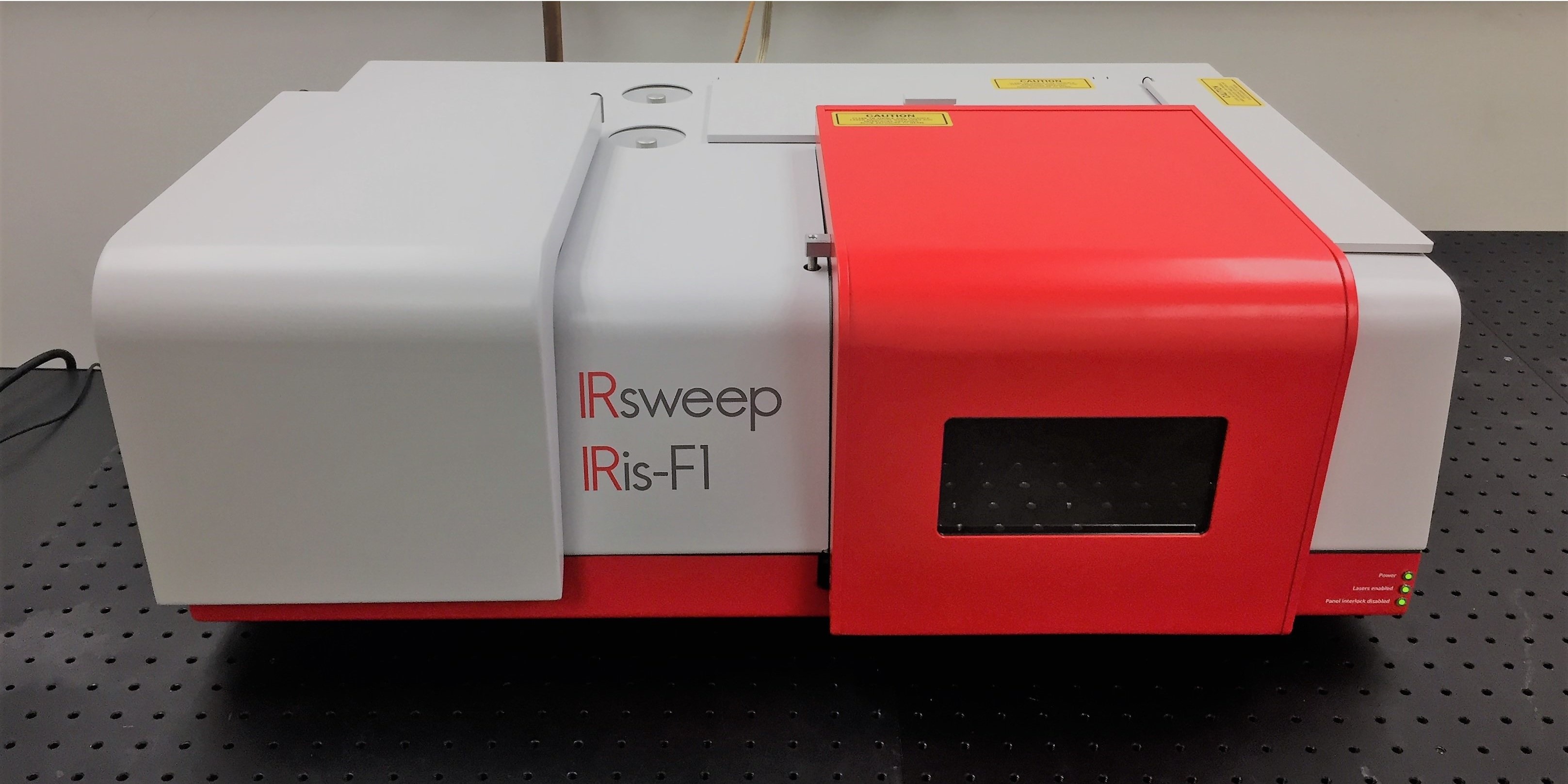
IRsweep IRis-F1 Spectrometer
## Techniques
Time-resolved spectroscopy with various sample geometries:
[**Transmission**](https://midir.lightsource.ca/mid-infrared-spectromicroscopy-mid-ir/transmission.md)
[**Reflection**](https://midir.lightsource.ca/mid-infrared-spectromicroscopy-mid-ir/reflection-transflection.md)
[**ATR**](https://midir.lightsource.ca/mid-infrared-spectromicroscopy-mid-ir/attenuated-total-reflection.md)
## Schematic
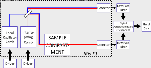
|---|---|
| Specifications ||
---
language: "en"
---
# Macro ATR Workflow Sample Change Procedure
## 1. Remove Sample
* Lower sample stage z all the way
* LOWER the microscope stage z by \~5000-6000 microns
**COLLISION DANGER:** Make sure the microscope z is **lowered** to prevent crystal arm colliding with objective
* Loosen crystal arm screws and swing away
* Rotate objective into front position
* Remove sample chuck
* Loosen left crystal arm screw until crystal arm can be removed
## 2. Clean Germanium Crystal and Sample Chuck
*Tip:* Careful when handling the crystal arm: **Never** touch the top surface of the ATR crystal. Clean the sample tip (bottom) with **lens paper only** . Contact your beamline staff if you have questions about the appropriate solvent to use for your system. **ONLY use water/alcohols.**
* Carefully clean the crystal tip and side with water/isopropanol (depending on sample). Finish with methanol and let completely dry before assembly
* Meanwhile, remove sample from chuck. Clean if needed and place new sample.
## 3. Reassemble Macro-ATR Plate and Load New Sample
* Reassemble crystal arm onto the macro-ATR plate in the swung out position with left screw fastened
From here on load your next sample and continue with another [Macro-ATR measurement](https://midir.lightsource.ca/mid-infrared-spectromicroscopy-mid-ir/bruker-operation-macro-atr.md).
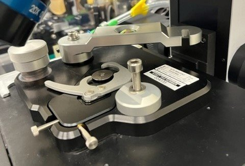
---
language: "en"
---
# Mail-in
## How the Mail-in Program Works:
In the mail-in program at Mid-IR the user prepares the sample and mails it to the Mid-IR beamline. Data collection is run by beamline staff.
Two pathways
1. Mail-in general users program
For the *general user program mail-in,* the user designs the experiment, [++mails++](https://midir.lightsource.ca/mid-infrared-spectromicroscopy-mid-ir/shipping.md) samples and analyzes the data. It is the user's responsibility to ensure the [++samples++](https://midir.lightsource.ca/mid-infrared-spectromicroscopy-mid-ir/samples.md) are appropriate for the desired measurements, the measurement parameters are determined before beam time and the user is available for communication during the beam time.
2. Mail-in as a fee based [++service++](https://www.lightsource.ca/industry/industry-services.php)
For the *fee based service* , the user has the choice of level of involvement and the fee for the service will reflect that. If you are interested, please [++contact++](https://midir.lightsource.ca/mid-infrared-spectromicroscopy-mid-ir/contact.md) us or [inquire with our Industry Services team](mailto:erika.bergen@lightsource.ca) to find out more information!
### Communicating During Beamtime
Beamline staff will work with your samples and collect data at the determined regions of interest with previously decided parameters. The user should be available as much as possible to answer questions and help staff manage the experimental run.
There are many options for communication. Please make a plan with beamline staff before your beam time for communication method.
1. Microsoft Teams (preferred)
2. phone/text
3. email
4. other, specify.
### Preparing samples
As much of the sample preparation as possible should be done at the user's home institution. Consideration should be made to insure the sample is stored securely and isn't damaged during shipment. The beamline staff are available to do sample mounting and other basic sample preparation. Preparation requirements should be agreed upon well before beam time and detailed instructions provided.
Information for general preparation of sample suitable for infrared measurements can be found in the [++sample++](https://midir.lightsource.ca/mid-infrared-spectromicroscopy-mid-ir/samples.md) section of the user guide.
For mail-in experiments it is important there is a fiduciary mark or an identifying feature on the sample that can be used for sample alignment, size determination, and locating the predetermined regions of interest.
### Experiment planning
Since you, the user, ***will not*** be the one controlling the instrument, it is important to make a detailed experimental plan before your beam time. This should detailed information about each sample, a priority ranking if you have more than one sample / region on interest. Parameters including, objective, scan size, number of scans and spectral resolution will all need to be determined prior to beam time.
**Please** [++**connect**++](https://midir.lightsource.ca/mid-infrared-spectromicroscopy-mid-ir/contact.md)**with beamline staff before your beam time to discuss your plan and confirm its feasibility.**
---
language: "en"
---
# Measurement Types
Our microscopes can be situated in the following measurement modes:
* [Transmission](https://midir.lightsource.ca/mid-infrared-spectromicroscopy-mid-ir/transmission.md)
* [Reflection/Transflection](https://midir.lightsource.ca/mid-infrared-spectromicroscopy-mid-ir/reflection-transflection.md)
* [Attenuated Total Reflection](https://midir.lightsource.ca/mid-infrared-spectromicroscopy-mid-ir/attenuated-total-reflection.md)
* [Sample Environments](https://midir.lightsource.ca/mid-infrared-spectromicroscopy-mid-ir/sample-environments.md)
---
language: "en"
---
# Mid-IR Beamline Documents
| File | Modified |
|---------------------------------------------------------------------------------------------------------------------------------------------------------------------------------------------------------------|--------------|
| [text_add-arrow.py](https://midir.lightsource.ca/__attachments/a_08407afc60b9248f412e56f96d539e1ef9ff83804a2f75a10d5b433f4cec739a/text_add-arrow.py.md?cb=3b4f6eeb5b219ab1d60844314126f6dd) | May 07, 2026 |
| [basic_1-3.py](https://midir.lightsource.ca/__attachments/a_32339b79af0d3235b7340bb653d91f6c589852019eb9abfbd324d22a5e093fca/basic_1-3.py.md?cb=f1fbce3d16a73b36086546f3067f6226) | May 07, 2026 |
| [basic2-3.py](https://midir.lightsource.ca/__attachments/a_4093c4c71a8305639dc178fc54241d35ce02400f78e9d7d488f23e1a063f3eb0/basic2-3.py.md?cb=9119d98a58ddf8dc12d0b21eaa0e8a94) | May 07, 2026 |
| [basic3-3.py](https://midir.lightsource.ca/__attachments/a_1c243865233afcfd1f0d187494f4d4b9bdc41a1f30126f6061623ac87ed5c1b0/basic3-3.py.md?cb=3c7fffcb498dd0c0bd198a6be509f721) | May 07, 2026 |
| [How_to_Submit_a_Beamtime_Request.pdf](https://midir.lightsource.ca/__attachments/a_1edb1ecab625d0f11cb5474fa94c20203680999932b5d20edfd6d66382655f70/How_to_Submit_a_Beamtime_Request.pdf.md?cb=9782d13bc18a5ec8e580ae226d7273a5) | May 07, 2026 |
| [hyperspectra_workflow.ows](https://midir.lightsource.ca/__attachments/a_a02e73ed6638ee514dc5d7a9fd5764e5aa96630ea21b21064b26a407f1e990bf/hyperspectra_workflow.ows.md?cb=4ab1edbae99213b2eed5ef180559f2ba) | May 07, 2026 |
| [matplotlib_tutorial.ows](https://midir.lightsource.ca/__attachments/a_96f850b6bcc4f5a6d3de93559de62fd369ccca0105a9cd9cdd3ca22ca5fa357a/matplotlib_tutorial.ows.md?cb=a0c89caca81d5b0abe3c10484437020f) | May 07, 2026 |
| [matplotlib_tutorial_OdfqT2P.ows](https://midir.lightsource.ca/__attachments/a_2d3b62fadfccc449fbd95ebb81cce55b2b762c70bbbbc9f468f309157e8499bd/matplotlib_tutorial_OdfqT2P.ows.md?cb=1617fbbc844d4cfadf7d671c1b88c4ed) | May 07, 2026 |
| [ROI_tutorial.ows](https://midir.lightsource.ca/__attachments/a_6fe331d87bc93d52350df9ec903f84dfb1819a9425db1636178164c6a41fcf49/ROI_tutorial.ows.md?cb=1b3a7d70322627642e0a9ce8e2eb187b) | May 07, 2026 |
| [110-file-and-data-table-widget-midir.ows](https://midir.lightsource.ca/__attachments/a_d2a643e8b07cacc3c82fc37e1b287e1bfc1d6088625a202fc90206e3eb2a75c7/110-file-and-data-table-widget-midir.ows.md?cb=075a2b0e10a7b79484f82b638b3f15a3) | May 07, 2026 |
---
language: "en"
---
# News
Beamline News
## Call for Proposals is Open!
### Scheduling Perion Jan-June 2027
The call for proposals is OPEN for beamtime in Cycle 43, January to June 2027. The call closes on Aug 19, 2026 at NOON Saskatoon time (CST).
Beamline contact:[**E-mail Scott Rosendahl, Mid-IR Beamline Responsible**](mailto:scott.rosendahl@lightsource.ca)
Learn more about [**Applying for Beamtime**](https://www.lightsource.ca/users/getting-started/applying-for-beamtime.php)and check out this[**Proposal Submission Webinar**](https://www.youtube.com/watch?v=q-M57H41xUg)video prepared by the USO.
### [→ See all Beamline News](https://midir.lightsource.ca/mid-infrared-spectromicroscopy-mid-ir/news.md)
REsearch Highlights
## FTIR Proves Effectiveness of a New and Improved Method for Cleaning Eggs
### Published: February, 2025
| Before hitting the shelves at your local grocery store, eggs roll through decontamination routines such as washing with hot water and drying - but sometimes these conventional methods damage egg quality. Researchers from the University of Saskatchewan use our [++Agilent++](https://midir.lightsource.ca/mid-infrared-spectromicroscopy-mid-ir/agilent.md) endstation and data collected with [++ATR-FTIR++](https://midir.lightsource.ca/mid-infrared-spectromicroscopy-mid-ir/attenuated-total-reflection.md) to investigate new eggshell washing practices. Additional Synchrotron X-ray micro-Computed Tomography (micro-CT) data was collected using the BioMedical Imaging and Therapy ([++BMIT++](https://bmit.lightsource.ca/)) beamline! In combination, these results prove the effectiveness of cold plasma, a non-thermal, chemical-free and eco-friendly technique for the decontamination of eggshells without sacrificing egg protein quality or eggshell cuticle coverage. **Read the paper!** Movasaghi, M.; Heydari, M.M.; Schwean-Lardner, K.; Kirychuk, S.; Thompson, B.; Zhang, L. Investigating cold plasma jet effectiveness for eggshell surface decontamination. *Food Control* **2025** , *168* , 110928. DOI: [++https://doi.org/10.1016/j.foodcont.2024.110928++](https://doi.org/10.1016/j.foodcont.2024.110928) | 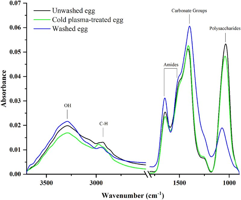 |
|---------------------------------------------------------------------------------------------------------------------------------------------------------------------------------------------------------------------------------------------------------------------------------------------------------------------------------------------------------------------------------------------------------------------------------------------------------------------------------------------------------------------------------------------------------------------------------------------------------------------------------------------------------------------------------------------------------------------------------------------------------------------------------------------------------------------------------------------------------------------------------------------------------------------------------------------------------------------------------------------------------------------------------------------------------------------------------------------------------------------------------------------------------------------------------------------------------------------------------------------------------------------------------------------|-------------------------------------------------------------------------------------------------------------------------------------------------------------------------------|
### [→ See all Research Highlights](https://midir.lightsource.ca/mid-infrared-spectromicroscopy-mid-ir/research-highlights.md)
* [Beamline News](https://midir.lightsource.ca/mid-infrared-spectromicroscopy-mid-ir/beamline-news.md)
* [Research Highlights](https://midir.lightsource.ca/mid-infrared-spectromicroscopy-mid-ir/research-highlights.md)
---
language: "en"
---
# NoMachine
This page provides information about remote control of the Mid-IR beamline workstations with NoMachine.
## Connecting to the instrument computer
Connection to instrument computers are done using the software NoMachine. There are two ways to access NoMachine:
1. through a [web browser](https://midir.lightsource.ca/mid-infrared-spectromicroscopy-mid-ir/nomachine.md#Web-Browser) or
2. by installing the [NoMachine Enterprise Client.](https://midir.lightsource.ca/mid-infrared-spectromicroscopy-mid-ir/nomachine.md#Client)
### Web Browser
Visit the following webpage on your computer: [sra.lightsource.ca](https://sra.lightsource.ca/)
* Enter the credentials supplied by the beamline staff:
* **Username:** Provided `nm-` account
* **Password:** Corresponding password
Continue through the remaining prompts by selecting **OK** and **Do Not Show Again** where applicable. Leave all default settings unchanged.
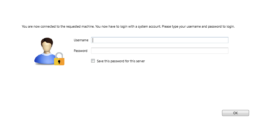
### NoMachine Client
#### Install NoMachine Enterprise Client:
Before beam time please download and install [++NoMachine Enterprise Client++](https://download.nomachine.com/enterprise/?product=enterprise-client)++:++
* On your PC, navigate to:
* Follow the download instructions for the NoMachine Enterprise Client appropriate to your computer and operating system
* Install the Client onto your computer
#### Setup your connection:
* Start NoMachine Enterprise Client
* Select Add under machines → Add connection
* Set connection settings:
* **Name:**your choice
* **Host Address:** `sra.lightsource.ca`
* Port Number: `4000`
* Protocol: `NX`
* Uncheck the **UDP** box and click **Add**
* If prompted about proxy settings, choose **Don't use a proxy** (unless you are on a proxied network), Continue
#### Logging into the Beamline Computer:
* Double-click the new connection to launch it.
* Enter **Username and Password\*\***
* Enter the `nm-` username and password provided by your beamline staff.
* Accept the presented certificate fingerprint.
* Double-Click the new connection to launch it.
* Username/Password: The `nm-` user and password.
* Click **OK** to all prompts and **Do Not Show Again** -- do not adjust any settings.
**\*\***At the beginning of your beamtime you will be provided with the instrument username and a password enabling remote access for the duration of your beamtime.
---
language: "en"
---
# Preparing a Proposal
Welcome! And thanks for deciding to submit a proposal for the Mid-IR beamline. On this page you'll find some tips on how to prepare a proposal for beamtime at the [++CLS++](https://www.lightsource.ca/users/getting-started/applying-for-beamtime.php). Please[++contact beamline staff++](https://midir.lightsource.ca/mid-infrared-spectromicroscopy-mid-ir/contact.md) if you are planning on submitting a proposal and we can help!
A successful proposal will be novel research and the aim of the project will be clearly identified. Explain the importance of the project and the reason why measurements at the CLS are required.
Please visit the [++About Us++](https://midir.lightsource.ca/mid-infrared-spectromicroscopy-mid-ir/about-us.md) page for details on the Mid-IR endstations:
The [**Bruker endstation**](https://midir.lightsource.ca/mid-infrared-spectromicroscopy-mid-ir/bruker.md) is optimized for high-brightness, diffraction-limited spectromicroscopy with synchrotron infrared radiation.
The [**Agilent endstation**](https://midir.lightsource.ca/mid-infrared-spectromicroscopy-mid-ir/agilent.md) is optimized for high spatial resolution, large field of view chemical infrared imaging.
The [**IRsweep endstation**](https://midir.lightsource.ca/mid-infrared-spectromicroscopy-mid-ir/irsweep.md) is optimized for high signal-to-noise, time-resolved spectroscopy measurements with microsecond time resolution.
## Getting Started
Step 1: [++Check++](https://www.lightsource.ca/users/getting-started/applying-for-beamtime.php) to see when the next call for proposals starts.
Step 2: Create a user account. Complete the registration form by clicking [++New user?++](http://user-portal.lightsource.ca/) on the log in page of the User Portal
Step 3: Create a new proposal. Click the + Create a Proposal button. Select General User Access
Step 4: The proposal
Step 5: Submission and results.
### For a short tutorial visit [++How to Submit a Proposal++](https://www.lightsource.ca/users/getting-started/user-portal-guide.php#HowtoSubmitaProposal)
The last Call for Proposals closed on **February 25, 2026.** The call results will be announced the week of **May 4, 2026.**
Results will be used for scheduling beamtime in **Cycle 42** (July-December, 2026). See the [++current schedule++](https://www.lightsource.ca/users/operations-schedule.php) for further information.
Stay tuned: The **next Call for Proposals** is expected in the **Fall of 2026**.
## The Proposal
Below is an outline of the type of material to prepare for your proposal. **The proposal is entirely created in the** [++**User Portal.**++](https://user-portal.lightsource.ca/) *It is recommended to draft the proposal in a word processing document and then enter into the user portal system.*
The proposal has a few main sections
1. **Description**
1. Abstract (200 words)
2. Research Area and Keywords
3. Scientific Description (800 words max): this part is really important, here you want to state the scientific problem, the current status of the research problem, and how this project at CLS will address the problem. Include specifics about what you plan on studying at the beamline, including something information about your sample
4. Societal, Economic and industrial Relevance (200 words max)
2. **Research Team**
1. Who is on the team, experience and publications related to the project proposal. (1000 words max plus one additional attachment)
3. **Beamlines** - In this section you outline your specific requirements at the CLS. Please talk to [++beamline staff++](https://midir.lightsource.ca/mid-infrared-spectromicroscopy-mid-ir/contact.md) as you prepare this section!
1. Facility requirements
2. Experimental procedure -- how will you prepare your samples, collect data and analyze data.
3. Justification of suitability
4. **Materials**
1. List of [samples](https://midir.lightsource.ca/mid-infrared-spectromicroscopy-mid-ir/samples.md) -- include details on the maximum number of samples you plan using in the proposal
2. Sample handling procedure (500 words)
3. Waste generation
Appendix: Attachment with preliminary results from previous CLS study or proof of concept or other lab analysis
## The details for Mid-IR
### Instruments:
Please visit the [++About Us++](https://midir.lightsource.ca/mid-infrared-spectromicroscopy-mid-ir/about-us.md) page for details on the Mid-IR endstations:
The [**Bruker endstation**](https://midir.lightsource.ca/mid-infrared-spectromicroscopy-mid-ir/bruker.md) is optimized for high-brightness, diffraction-limited spectromicroscopy with synchrotron infrared radiation.
The [**Agilent endstation**](https://midir.lightsource.ca/mid-infrared-spectromicroscopy-mid-ir/agilent.md) is optimized for high spatial resolution, large field of view chemical infrared imaging.
The [**IRsweep endstation**](https://midir.lightsource.ca/mid-infrared-spectromicroscopy-mid-ir/irsweep.md) is optimized for high signal-to-noise, time-resolved spectroscopy measurements with microsecond time resolution.
### Techniques:
In the user portal, the mid-IR beamline has the following four techniques listed. Beside each technique is the type of measurement
* **FTIR spectromicroscopy** - Measurements using the single-point detector. For example point-by-point mapping or single-point microscopic measurements
* **Chemical Infrared Imaging -** Array (FPA) Imaging
* **ATR infrared spectroscopy -** Measurement in ATR configuration.
* **Fourier Transform Infrared Spectroscopy (FTIR)** - bulk spectroscopic measurement
### TIPS
[++Contact beamline staff++](https://midir.lightsource.ca/mid-infrared-spectromicroscopy-mid-ir/contact.md) before submitting a proposal!
Clearly state your research problem so a non-expert can understand
Prepare a detailed experimental plan
Demonstrate a clear path for publication of the CLS results
## Cross Beamline
**Multiple Beamline Proposal** - If a proposal is to be performed on multiple beamlines, you can add additional Facilities to the same proposal.
## Submit and Notifications
When you submit your proposal you and all your team members will get an email notification that a proposal has been successfully submitted. After submitting, you will get emails for items relating to your proposal that need your attention. Notifications can include:
**Review Required (Safety, Technical or Peer Review)**
**Clarification of Issues (Safety, Technical or Peer Review) during the review process**
**Responses to Clarification of Issues during the review process**
**Permit Amendments -- notification of review status (Approved/Denied)**
---
language: "en"
---
# Publications
Don't see your publications here? We would love to celebrate your accomplishments! Let us know when data you've collected at the Mid-IR beamline contributes to publishing research articles, submitting your thesis or presenting at conferences and events to be featured on our website or at the CLS Annual Users' Meeting. Make sure you [report your published works](https://www.lightsource.ca/users/your-cls-experiment/after-your-beamtime.php#ReportingPublications) to the CLS, too!
| Cite | Date |
|---------------------------------------------------------------------------------------------------------------------------------------------------------------------------------------------------------------------------------------------------------------------------------------------------------------------------------------------------------------------------------------------------------------------------------------------------------------------------------------------------|------------|
| Black, Tallan; Boseley, Rhiannon E.; Quirk, Amanda; Young, Kaylen M.; Lunardi-Baccetto, Sarah et al. (2026). *Adult Rat Offspring Exposed to THC during Gestation Exhibit Distinct Biomolecular Changes Identified by X-ray Fluorescence Imaging and Fourier Transform Infrared Spectroscopy in Cortico-Limbic Circuits* . ACS Chemical Neuroscience . [10.1021/acschemneuro.5c00752](https://dx.doi.org/10.1021/acschemneuro.5c00752). | 2026-02-09 |
| Deng, Tianyang (2026). *Developing Bioanalytical Platforms for Studying Cyanobacteria with Vibrational Spectroscopy and Hyperspectral Imaging* . Supervisor: Greener, Jesse; Vincent, Warwick. Quebec, Canada: Université Laval. . | 2026-02-16 |
| Hillary Soita (2026). *MID IR Beamline Publications Testing*. Supervisor: Eshi. SK: CLS. . | 2026-05-07 |
| Houseman, Melissa (2026). *Time-Resolved IR Spectroelectrochemistry of a Surface Confined Redox Couple* . Supervisor: Burgess, Ian. Saskatchewan, Canada: University of Saskatchewan. . | 2026-03-23 |
| Pensini, Erica; Santillan, Ana Sofia Pina; dalla Pasqua, Maya; Truong, Kenneth; Marangoni, Alejandro G. et al. (2026). *From miscibility to mesoscale heterogeneity: tannic-acid-mediated assembly of pyridine in aqueous liquids* . Soft Matter . [10.1039/d6sm00605a](https://dx.doi.org/10.1039/d6sm00605a). | 2026-01-01 |
| Santillan, Ana Sofia Pina; dalla Pasqua, Maya; Marangoni, Alejandro G.; Truong, Kenneth; Pensini, Erica et al. (2026). *Sulfolane modulates tannic acid--pyridine organization in bulk water and at a silica--water interface* . Colloids and Surfaces C Environmental Aspects 4, 100105. [10.1016/j.colsuc.2026.100105](https://dx.doi.org/10.1016/j.colsuc.2026.100105). | 2026-11-01 |
| Song, Xing; Cao, Yiqi; Zhang, Baiyu; Mesburis, Rebecca; Rosendahl, Scott et al. (2026). *Disposable Face Masks and PBDEs in Aquatic Environments: Co-contamination and Impacts* . Water Research , 125874. [10.1016/j.watres.2026.125874](https://dx.doi.org/10.1016/j.watres.2026.125874). | 2026-04-03 |
| Tu, Jiangying; Gossen, Bruce D.; Tu, Kaiyang; Stobbs, Jarvis; Reid, Joel W. et al. (2026). *In planta storage of boron and calcium affects clubroot severity in Brassica napus* . Plant Physiology and Biochemistry 238, 111604. [10.1016/j.plaphy.2026.111604](https://dx.doi.org/10.1016/j.plaphy.2026.111604). | 2026-09-01 |
| Yang, Min; Chen, Bing; Kang, Qiao; Wang, Anran; Yuan, Runbo et al. (2026). *Prediction of microplastic transport in oil-contaminated waters* . Marine Pollution Bulletin 222, 118795. [10.1016/j.marpolbul.2025.118795](https://dx.doi.org/10.1016/j.marpolbul.2025.118795). | 2026-01-01 |
| Ashe, Paula; Tu, Kaiyang; Stobbs, Jarvis A.; Dynes, James J.; Vu, Miranda et al. (2025). *Applications of synchrotron light in seed research: an array of x-ray and infrared imaging methodologies* . Frontiers in Plant Science 15. [10.3389/fpls.2024.1395952](https://dx.doi.org/10.3389/fpls.2024.1395952). | 2025-02-17 |
| Burgess, Ian J.; Morhart, Tyler A.; Flaman, Grace T.; Boyle, Nicole D.; Deng, Tiangyang et al. (2025). *Into the Groove: Analytical Applications of ATR-FTIR Microstructured Internal Reflection Elements* . Analytical Chemistry . [10.1021/acs.analchem.4c05431](https://dx.doi.org/10.1021/acs.analchem.4c05431). | 2025-05-01 |
| Coker, Oluwafemi Jeremiah (2025). *Development of Hybrid Meat Products by Replacing Animal Fats with Faba bean Protein Isolate-Stabilized Canola Oil-In-Water Emulsion Gel* . Supervisor: Shand, Phyllis J; Ghosh, Supratim. Saskatchewan, Canada: University of Saskatchewan. . | 2025-11-03 |
| Dobson, S.; Marangoni, A.G. (2025). *Evaluating the effect of plant protein functionalities on the performance of high-protein plant-based cheese* . Food Chemistry 492, 145553. [10.1016/j.foodchem.2025.145553](https://dx.doi.org/10.1016/j.foodchem.2025.145553). | 2025-11-01 |
| Goff, Kira L.; Wilson, Kenneth E.; Ellis, Thomas H. (2025). *Using synchrotron FTIR spectromicroscopy to assess physiological and metabolic changes in photosynthetic mutants of Chlamydomonas reinhardtii* . Canadian Journal of Chemistry . [10.1139/cjc-2024-0237](https://dx.doi.org/10.1139/cjc-2024-0237). | 2025-04-24 |
| Indore, Navnath S.; Jayas, Digvir S.; Karunakaran, Chithra; Stobbs, Jarvis; Bondici, Viorica F. et al. (2025). *Application of synchrotron imaging techniques for study of changes in microstructural and nutritional properties of different wheat classes in storage* . Journal of Stored Products Research 111, 102576. [10.1016/j.jspr.2025.102576](https://dx.doi.org/10.1016/j.jspr.2025.102576). | 2025-05-01 |
| Jiang, Yunfei; Lahlali, Rachid; Bueckert, Rosalind; Kumar, Saroj; Karunakaran, Chithra et al. (2025). *Unravelling Compositional Changes in Field Pea Leaf Cuticles Under Heat Stress Using Synchrotron‐Based Fourier Transform Mid‐Infrared Spectroscopy* . Journal of Agronomy and Crop Science 211(4) . [10.1111/jac.70099](https://dx.doi.org/10.1111/jac.70099). | 2025-07-01 |
| Li, Mengna; Huang, Guohe; Chen, Xiujuan; Xu, Zeyuan; Huang, Jing et al. (2025). *Development of an EOR-produced petroleum wastewater treatment system through integrated polyacrylonitrile membrane and ZrO2/sericin technologies: revelation of interactive mechanism based on synchrotron and XDLVO analyses* . npj Clean Water 8(1) . [10.1038/s41545-025-00454-6](https://dx.doi.org/10.1038/s41545-025-00454-6). | 2025-03-30 |
| Movasaghi, Mina; Heydari, Mohamad Mehdi; Schwean-Lardner, Karen; Kirychuk, Shelley; Thompson, Brooke et al. (2025). *Investigating cold plasma jet effectiveness for eggshell surface decontamination* . Food Control 168, 110928. [10.1016/j.foodcont.2024.110928](https://dx.doi.org/10.1016/j.foodcont.2024.110928). | 2025-02-01 |
| Mundboth, Kiran (2025). *Canadian Light Source -- 20 Years of Discovery* . Canadian Journal of Chemistry . [10.1139/cjc-2024-0278](https://dx.doi.org/10.1139/cjc-2024-0278). | 2025-05-06 |
| Njeru, Harriet K.; Knudsen, Knud E. Bach; Stobbs, Jarvis A.; Tu, Kaiyang; Woyengo, Tofuko A. et al. (2025). *Porcine *in vitro* digestion and matrix structure of undigested residue of xylanase‐ and cellulase‐supplemented maize and wheat* . Journal of the Science of Food and Agriculture . [10.1002/jsfa.14179](https://dx.doi.org/10.1002/jsfa.14179). | 2025-02-11 |
| Stacie Lynn Dobson (2025). *Development and Evaluation of High-Protein Plant-Based Cheese* . Supervisor: Marangoni , Alejandro. Guelph, Canada: The University of Guelph. . | 2025-05-21 |
| Stobbs, Jarvis A. (2025). *Multiscale Analysis Framework for Chocolate Tempering using Phospholipids* . Supervisor: Marangoni, Alejandro G.. Ontario, Canada: University of Guelph. . | 2025-05-09 |
| Stobbs, Jarvis A.; Ghazani, Saeed M.; Tu, Kaiyang; Pensini, Erica; Fameau, Anne-Laure et al. (2025). *Dimyristoylphosphoethanolamine Addition During Chocolate Manufacture Promotes Proper Tempering under Simple Cooling Conditions without Shear* . Crystal Growth and Design 25(12) , 4621-4635. [10.1021/acs.cgd.5c00575](https://dx.doi.org/10.1021/acs.cgd.5c00575). | 2025-06-04 |
| Tu, Jiangying; Karunakaran, Chithra; Wei, Yangdou; Peng, Gary (2025). *Barley leaf cell-wall responses to the penetration by adapted and nonadapted powdery mildew fungi revealed with the thermal-source based Fourier transform infrared microspectroscopy and focal plane array* . Canadian Journal of Plant Pathology , 1-14. [10.1080/07060661.2024.2441916](https://dx.doi.org/10.1080/07060661.2024.2441916). | 2025-01-13 |
| Wei, Wei; Du, Cheng; Ge, Jiawei; Wang, Xiang; Chen, Zuolong et al. (2025). *Bio‐Inspired Cascade Photocatalysis on Fe Single‐Atom Carbon Nitride Upcycles Plastic Wastes for Effective Acetic Acid Production* . Advanced Energy Materials . [10.1002/aenm.202505453](https://dx.doi.org/10.1002/aenm.202505453). | 2025-12-26 |
| Wu, Runqi; You, Xiang; Dey, Trinanjan; Flaman, Grace; Rosendahl, Scott et al. (2025). *Orthogonal Solvent Mediation for Solid Composite Electrolyte Synthesis: Phase Integration via Particle Dispersion* . Patent Number: [10.26434/chemrxiv-2025-0b2zx](https://patents.google.com/patent/10.26434/chemrxiv-2025-0b2zx/en). | 2025-11-17 |
| Bartokova, Bibiana; Marangoni, Alejandro G.; Pensini, Erica (2024). *Role of heads and tails on tetrahydrofuran- and dimethyl sulfoxide-water separation by glycerol and sucrose esters* . Physics of Fluids 36(7) . [10.1063/5.0209824](https://dx.doi.org/10.1063/5.0209824). | 2024-07-01 |
| Bibiana Bartokova (2024). *Impact of Amphiphiles, Water Chemistry and Co-contaminants on Miscible Contaminant Mixing Behaviour* . Supervisor: Pensini, E.. Ontario, Canada: University of Guelph. . | 2024-04-03 |
| Boseley, Rhiannon E.; Sylvain, Nicole J.; Peeling, Lissa; Kelly, Michael E.; Pushie, M. Jake et al. (2024). *A review of concepts and methods for FTIR imaging of biomarker changes in the post-stroke brain* . Biochimica et Biophysica Acta - Biomembranes 1866(3) , 184287. [10.1016/j.bbamem.2024.184287](https://dx.doi.org/10.1016/j.bbamem.2024.184287). | 2024-03-01 |
| Cao, Yiqi; Zhang, Baiyu; Song, Xing; Dong, Guihua; Zhang, Yuanmei et al. (2024). *Polyhydroxybutyrate Plastics Show Rapid Disintegration and More Straightforward Biogeochemical Impacts than Polyethylene under Marine Biofragmentation* . Environmental Science and Technology . [10.1021/acs.est.4c04639](https://dx.doi.org/10.1021/acs.est.4c04639). | 2024-07-24 |
| Dong, Shu; Xie, Geng; Xu, Shihong; Tan, Xuehai; Chaudhary, Madhusudan et al. (2024). *Cellulose-Encapsulated Composite Electrolyte Design: Toward Chemically and Mechanically Enhanced Solid-Sodium Batteries* . ACS Nano . [10.1021/acsnano.4c03910](https://dx.doi.org/10.1021/acsnano.4c03910). | 2024-06-12 |
| Elferjani, Raed; Pahari, Shankar; Soolanayakanahally, Raju; Ballantyne, Krista; Nambara, Eiji et al. (2024). *Drought induced metabolic shifts and water loss mechanisms in canola: role of cysteine, phenylalanine and aspartic acid* . Frontiers in Plant Science 15. [10.3389/fpls.2024.1385414](https://dx.doi.org/10.3389/fpls.2024.1385414). | 2024-12-23 |
| Grace T. Flaman (2024). *Infrared Chemical Imaging of Custom-Made Microfluidic Devices* . Supervisor: Burgess, Ian J.. Saskatchewan, Canada: University of Saskatchewan. . | 2024-01-05 |
| Guevara, Victor; Nagy, Carlene; Yang, Jen-Chieh; He, Jiangfeng; E. Rodriguez-Espinosa, Maria et al. (2024). *Using Conventional Ruminant Techniques and Molecular Spectroscopy to Study the Impact of Additive Fibrolytic Enzymes and Maturity Stage on Nutritional and Molecular Structural Changes of Legume and Legume-Cereal Intercropped Silage* . . [10.5772/intechopen.114241](https://dx.doi.org/10.5772/intechopen.114241). | 2024-02-20 |
| Indore, Navnath S. (2024). *Elucidation of changes in food grains due to spoilage in bulk storage using advanced imaging for post-harvest management* . Supervisor: Jayas, Digvir; Karunakaran, Chithra. Manitoba, Canada: University of Manitoba. . | 2024-01-15 |
| Ketki Srivastava (2024). *Everything, Everywhere, All At Once: Micro- and Nano-Fabrication for Sensitive, Homogenous and Spatio-Temporally-Resolved Raman and Infrared Spectroscopy Sensors* . Supervisor: Odijk, M.; van den Berg, A.. Enschede, The Netherlands: University of Twente. . | 2024-09-20 |
| Li, Mengna; Huang, Guohe; Chen, Xiujuan; Xiao, Huining; An, Chunjiang et al. (2024). *Development of an antimicrobial and antifouling PES membrane with ZnO/poly(hexamethylene biguanide) nanocomposites incorporation* . Chemical Engineering Journal 481, 148744. [10.1016/j.cej.2024.148744](https://dx.doi.org/10.1016/j.cej.2024.148744). | 2024-02-01 |
| Nicole D. K. Boyle (2024). *Pairing Synchrotron Radiation with an ATR-Integrated Microreactor for In-Situ Spatiotemporal Characterization of Chemical Reactions* . Supervisor: Burgess, Ian, J.. Saskatchewan, Canada: University of Saskatchewan. . | 2024-01-17 |
| Pushie, M. Jake; Sylvain, Nicole J.; Hou, Huishu; George, Dominic; Kelly, Michael E. et al. (2024). *Ion Dyshomeostasis in the Early Hyperacute Phase after a Temporary Large-Vessel Occlusion Stroke* . ACS Chemical Neuroscience . [10.1021/acschemneuro.3c00685](https://dx.doi.org/10.1021/acschemneuro.3c00685). | 2024-05-14 |
| Pushie, M Jake; Sylvain, Nicole J; Hou, Huishu; Pendleton, Nicole; Wang, Richard et al. (2024). *X-ray fluorescence mapping of brain tissue reveals the profound extent of trace element dysregulation in stroke pathophysiology* . Metallomics . [10.1093/mtomcs/mfae054](https://dx.doi.org/10.1093/mtomcs/mfae054). | 2024-11-15 |
| Shen, Jian; Huang, Guohe; Yao, Yao; Li, Mengna; Zhang, Peng et al. (2024). *Development of calcium-modified biochar for enhanced phytoremediation of human-induced salt pollutants (HISPs)* . Chemosphere 355, 141860. [10.1016/j.chemosphere.2024.141860](https://dx.doi.org/10.1016/j.chemosphere.2024.141860). | 2024-05-01 |
| Shen, Jian; Huang, Guohe; Yao, Yao; Zhang, Peng; Rosendahl, Scott et al. (2024). *Surface Alteration on Biochar in Long-Term Application: Insights into Pyrolysis, Freeze-Thaw Aging, and Dissipation* . Surfaces and Interfaces , 104118. [10.1016/j.surfin.2024.104118](https://dx.doi.org/10.1016/j.surfin.2024.104118). | 2024-03-01 |
| Shen, Jian; Huang, Guohe; Yao, Yao; Zhang, Peng; Rosendahl, Scott et al. (2024). *Thermodynamic assessment of Tetrabromobisphenol A (TBBPA) persistence over time in freeze-thaw aged biochar and its environmental significance* . Industrial Crops and Products 221, 119255. [10.1016/j.indcrop.2024.119255](https://dx.doi.org/10.1016/j.indcrop.2024.119255). | 2024-07-31 |
| Sivakumar, Chitra; Stobbs, Jarvis A.; Tu, Kaiyang; Karunakaran, Chithra; Paliwal, Jitendra et al. (2024). *Investigating the microstructure of chickpea and navy bean flour blends produced by roller milling: Insights from Fourier transform mid-infrared spectroscopy, scanning electron microscopy and synchrotron X-ray techniques* . Advanced Powder Technology 35(11) , 104674. [10.1016/j.apt.2024.104674](https://dx.doi.org/10.1016/j.apt.2024.104674). | 2024-11-01 |
| Sivakumar, Chitra; Stobbs, Jarvis A.; Tu, Kaiyang; Karunakaran, Chithra; Paliwal, Jitendra et al. (2024). *Investigating the particle morphology and molecular structure of roller-milled yellow pea flours using high-resolution imaging, mid-infrared spectroscopy, and synchrotron X-rays* . Journal of Food Measurement and Characterization . [10.1007/s11694-024-03082-9](https://dx.doi.org/10.1007/s11694-024-03082-9). | 2024-12-30 |
| Stobbs, Jarvis A.; Pensini, Erica; Ghazani, Saeed M.; Leontowich, Adam F. G.; Quirk, Amanda et al. (2024). *Phospholipid Self-Assembly in Cocoa Butter Provides a Crystallizing Surface for Seeding the Form V Polymorph in Chocolate* . Crystal Growth and Design . [10.1021/acs.cgd.3c01130](https://dx.doi.org/10.1021/acs.cgd.3c01130). | 2024-03-21 |
| Tu, Jiangying; Qin, Li; Karunakaran, Chithra; Wei, Yangdou; Peng, Gary et al. (2024). *Lignin accumulation in cell wall plays a role in clubroot resistance* . Frontiers in Plant Science 15. [10.3389/fpls.2024.1401265](https://dx.doi.org/10.3389/fpls.2024.1401265). | 2024-07-23 |
| Wang, Hongjie; Chen, Xiujuan; Chen, Bing; Zhao, Yuming; Zhang, Baiyu et al. (2024). *Development of a spiropyran-assisted cellulose aerogel with switchable wettability as oil sorbent for oil spill cleanup* . Science of the Total Environment , 171451. [10.1016/j.scitotenv.2024.171451](https://dx.doi.org/10.1016/j.scitotenv.2024.171451). | 2024-03-01 |
| Yin, Jianan (2024). *Nanocellulose-based materials for sustainable soil remediation and water purification* . Supervisor: Huang, Guohe. Saskatchewan, Canada: University of Regina. . | 2024-01-31 |
| Atkinson, Noah; Morhart, Tyler A.; Wells, Garth; Flaman, Grace T.; Petro, Eric et al. (2023). *Microfabrication Process Development for a Polymer-Based Lab-on-Chip Concept Applied in Attenuated Total Reflection Fourier Transform Infrared Spectroelectrochemistry* . Sensors 23(14) , 6251. [10.3390/s23146251](https://dx.doi.org/10.3390/s23146251). | 2023-07-08 |
| Bartokova, Bibiana; Marangoni, Alejandro G.; Laredo, Thamara; Pensini, Erica (2023). *Effect of sorbitan ester structure on the separation between tetrahydrofuran and water* . Frontiers in Soft Matter 3. [10.3389/frsfm.2023.1329058](https://dx.doi.org/10.3389/frsfm.2023.1329058). | 2023-12-18 |
| Cai, Mengfan; Qi, Zhiming; Guy, Christophe; An, Chunjiang; Chen, Xiujuan et al. (2023). *Insights into the abiotic fragmentation of biodegradable mulches under accelerated weathering conditions* . Journal of Hazardous Materials 454, 131477. [10.1016/j.jhazmat.2023.131477](https://dx.doi.org/10.1016/j.jhazmat.2023.131477). | 2023-07-01 |
| Dobson, S.; Stobbs, J.; Laredo, T.; Marangoni, A.G. (2023). *A facile strategy for plant protein fiber formation without extrusion or shear processing* . Innovative Food Science and Emerging Technologies 86, 103385. [10.1016/j.ifset.2023.103385](https://dx.doi.org/10.1016/j.ifset.2023.103385). | 2023-06-01 |
| Flaman, Grace T.; Boyle, Nicole D.; Vermelle, Cyprien; Morhart, Tyler A.; Ramaswami, Bdhanya et al. (2023). *Chemical Imaging of Mass Transport Near the No-Slip Interface of a Microfluidic Device using Attenuated Total Reflection--Fourier Transform Infrared Spectroscopy* . Analytical Chemistry 95(11) , 4940-4949. [10.1021/acs.analchem.2c04880](https://dx.doi.org/10.1021/acs.analchem.2c04880). | 2023-03-07 |
| Gao, Sichen; Huang, Gordon; Zhang, Peng; Xin, Xiaying; Yin, Jianan et al. (2023). *Toxicity and mechanism of nanoplastics to phytoplankton in high-latitude aquatic ecosystems of Canadian prairie: Effects of multiple environmental factors* . Science of the Total Environment 893, 164676. [10.1016/j.scitotenv.2023.164676](https://dx.doi.org/10.1016/j.scitotenv.2023.164676). | 2023-10-01 |
| Gao, Sichen; Huang, Gordon; Zhang, Peng; Xin, Xiaying; Yin, Jianan et al. (2023). *Rethinking the effects of micro/nanoplastics from the global environmental change and systematic perspective: An aquatic environmental system-based comprehensive assessment approach of micro/nanoplastic impacts* . Journal of Hazardous Materials 457, 131695. [10.1016/j.jhazmat.2023.131695](https://dx.doi.org/10.1016/j.jhazmat.2023.131695). | 2023-09-01 |
| Grossutti, Michael; D'Amico, Joseph; Quintal, Jonathan; MacFarlane, Hugh; Wareham, W. Callum et al. (2023). *Deep Generative Modeling of Infrared Images Provides Signature of Cracking in Cross-Linked Polyethylene Pipe* . ACS Applied Materials and Interfaces 15(18) , 22532-22542. [10.1021/acsami.3c02564](https://dx.doi.org/10.1021/acsami.3c02564). | 2023-04-25 |
| Indore, Navnath S.; Jayas, Digvir S.; Karunakaran, Chithra; Stobbs, Jarvis; Bondici, Viorica F. et al. (2023). *Study of Microstructural, Nutritional, and Biochemical Changes in Hulled and Hulless Barley during Storage Using X-ray and Infrared Techniques* . Foods 12(21) , 3935. [10.3390/foods12213935](https://dx.doi.org/10.3390/foods12213935). | 2023-10-27 |
| Indore, Navnath S.; Karunakaran, Chithra; Jayas, Digvir S.; Bondici, Viorica F.; Vu, Miranda et al. (2023). *Mapping biochemical and nutritional changes in durum wheat due to spoilage during storage* . Heliyon 9(11) , e22139. [10.1016/j.heliyon.2023.e22139](https://dx.doi.org/10.1016/j.heliyon.2023.e22139). | 2023-11-01 |
| Laing, Emma; Stone, Andrea K.; Shi, Dai; Pickard, Mark; House, James D. et al. (2023). *Effect of infrared heating on the nutritional properties of yellow pea and green lentil flours* . Cereal Chemistry 100(3) , 614-627. [10.1002/cche.10653](https://dx.doi.org/10.1002/cche.10653). | 2023-02-03 |
| Lyu, Linxiang; Wang, Zheng; Bagchi, Monisha; Ye, Zhibin; Soliman, Ahmed et al. (2023). *An investigation into the aging of disposable face masks in landfill leachate* . Journal of Hazardous Materials 446, 130671. [10.1016/j.jhazmat.2022.130671](https://dx.doi.org/10.1016/j.jhazmat.2022.130671). | 2023-03-01 |
| Mitchell, Jerit Leo (2023). *Unprecedented preservation of soft tissues and organic matter in fossils characterized by synchrotron radiation techniques* . Supervisor: Barbi, Mauricio; McKeller, Ryan. Saskatchewan, Canada: University of Regina. . | 2023-11-20 |
| Mitchell, Jerit L.; McKellar, Ryan C.; Barbi, Mauricio; Coulson, Ian M.; Bukejs, Andris et al. (2023). *Morphological and organic spectroscopic studies of a 44-million-year-old leaf beetle (Coleoptera: Chrysomelidae) in amber with endogenous remains of chitin* . Scientific Reports 13(1) , 5876. [10.1038/s41598-023-32557-w](https://dx.doi.org/10.1038/s41598-023-32557-w). | 2023-04-11 |
| Najib, Tahereh (2023). *Microwave-infrared/vacuum drying, roasting, and convective air drying of germinated lentils to modify physicochemical and functional properties* . Supervisor: Meda, Venkatesh. Saskatchewan, Canada: University of Saskatchewan. . | 2023-08-08 |
| Najib, Tahereh; Heydari, Mohamad Mehdi; Tu, Kaiyang; Meda, Venkatesh (2023). *Modification in starch structure of soaked and germinated lentil seeds under various thermal processing methods, including conventional, microwave, and microwave-assisted techniques* . Food Chemistry Advances 2, 100267. [10.1016/j.focha.2023.100267](https://dx.doi.org/10.1016/j.focha.2023.100267). | 2023-10-01 |
| Najib, Tahereh; Heydari, Mohamad Mehdi; Tu, Kaiyang; Vu, Miranda; Meda, Venkatesh et al. (2023). *Protein structural changes in lentil flour during soaking/germination and thermal treatments: Indication of nutritional and functional properties* . Food Chemistry Advances 3, 100475. [10.1016/j.focha.2023.100475](https://dx.doi.org/10.1016/j.focha.2023.100475). | 2023-12-01 |
| Patel, Vedant; Marangoni, Alejandro G.; Ghazani, Saeed Mirzaee; Laredo, Thamara; Stobbs, Jarvis et al. (2023). *Effect of bacterial surfactants on the phase behavior of miscible pollutants in water* . Colloids and Surfaces C Environmental Aspects 1, 100013. [10.1016/j.colsuc.2023.100013](https://dx.doi.org/10.1016/j.colsuc.2023.100013). | 2023-11-01 |
| Rengyu Yue (2023). *Advancing Shoreline Oil Spill Response with Eco-friendly Functional Nanomaterials* . Supervisor: An, Chunjiang; Ye, Zhibin. Quebec, Canada: Concordia University. . | 2023-11-28 |
| Shabtai, Itamar A.; Wilhelm, Roland C.; Schweizer, Steffen A.; Höschen, Carmen; Buckley, Daniel H. et al. (2023). *Calcium promotes persistent soil organic matter by altering microbial transformation of plant litter* . Nature Communications 14(1) . [10.1038/s41467-023-42291-6](https://dx.doi.org/10.1038/s41467-023-42291-6). | 2023-10-19 |
| Sivakumar, Chitra; Stobbs, Jarvis A.; Tu, Kaiyang; Karunakaran, Chithra; Paliwal, Jitendra et al. (2023). *Molecular characterization of green lentil flours using synchrotron X-rays and Fourier transform mid-infrared techniques* . Powder Technology 426, 118674. [10.1016/j.powtec.2023.118674](https://dx.doi.org/10.1016/j.powtec.2023.118674). | 2023-08-01 |
| Srivastava, K.; Boyle, N. D.; Flaman, G. T.; Ramaswami, B.; van den Berg, A. et al. (2023). **In situ* spatiotemporal characterization and analysis of chemical reactions using an ATR-integrated microfluidic reactor* . Lab on a Chip . [10.1039/d3lc00521f](https://dx.doi.org/10.1039/d3lc00521f). | 2023-01-01 |
| Stephen Campbell (2023). *Vibrational spectroscopy and the structure of solids: The example of carbonate minerals* . Supervisor: Poduska, Kristin, M.. Newfoundland and Labrador, Canada: Memorial University. . | 2023-08-01 |
| Yang, Min; Zhang, Baiyu; Chen, Xiujuan; Kang, Qiao; Gao, Boyang et al. (2023). *Transport of Microplastic and Dispersed Oil Co-contaminants in the Marine Environment* . Environmental Science and Technology 57(14) , 5633-5645. [10.1021/acs.est.2c08716](https://dx.doi.org/10.1021/acs.est.2c08716). | 2023-03-27 |
| Yin, Jianan; Huang, Guohe; Chen, Xiujuan; Shen, Jian; An, Chunjiang et al. (2023). *Recyclable sericin/nanocellulose aerogel with high adsorption capacity for tetrabromobisphenol A in water: Insight from DFT calculations* . Chemical Engineering Journal 474, 145695. [10.1016/j.cej.2023.145695](https://dx.doi.org/10.1016/j.cej.2023.145695). | 2023-10-01 |
| Yin, Jianan; Huang, Guohe; Xiao, Huining; Chen, Ning; An, Chunjiang et al. (2023). *Bioinspired and dual-functional nanocellulose aerogels for water disinfection and heavy metal removal* . Nano Today 51, 101918. [10.1016/j.nantod.2023.101918](https://dx.doi.org/10.1016/j.nantod.2023.101918). | 2023-08-01 |
| Alivia Mukherjee (2022). *Generation of activated carbon from spent coffee grounds: Process optimization, kinetics and CO2 capture* . Supervisor: Ajay K. Dalai, Catherine Niu. Saskatchewan, Canada: University of Saskatchewan. . | 2022-09-15 |
| Chen, Xiujuan; Huang, Guohe; An, Chunjiang; Feng, Renfei; Wu, Yinghui et al. (2022). *Superwetting polyethersulfone membrane functionalized with ZrO2 nanoparticles for polycyclic aromatic hydrocarbon removal* . Journal of Materials Science and Technology 98, 14-25. [10.1016/j.jmst.2021.01.063](https://dx.doi.org/10.1016/j.jmst.2021.01.063). | 2022-01-01 |
| Chen, Xiujuan; Liu, Yunqiu; Huang, Gordon; An, Chunjiang; Feng, Renfei et al. (2022). *Functional flax fiber with UV-induced switchable wettability for multipurpose oil-water separation* . Frontiers of Environmental Science and Engineering 16(12) . [10.1007/s11783-022-1588-6](https://dx.doi.org/10.1007/s11783-022-1588-6). | 2022-06-20 |
| Deng, Ganqi; Nagy, Carlene; Yu, Peiqiang (2022). *Combined molecular spectroscopic techniques (SR-FTIR, XRF, ATR-FTIR) to study physiochemical and nutrient profiles of Avena sativa grain and nutrition and structure interactive association properties* . Critical Reviews in Food Science and Nutrition , 1-13. [10.1080/10408398.2022.2045470](https://dx.doi.org/10.1080/10408398.2022.2045470). | 2022-03-03 |
| Denis AB Therien (2022). *Plasmonically-Active Nanomaterials for Enhanced Second- Harmonic Generation and Chemical ReactionsHarmonic Generation and Chemical Reaction* . Supervisor: Lagugné-Labarthet, François. Ontario, Canada: The University of Western Ontario. . | 2022-04-13 |
| Earnden, Laura; Marangoni, Alejandro G.; Laredo, Thamara; Stobbs, Jarvis; Pensini, Erica et al. (2022). *Self-Assembled glycerol monooleate demixes miscible liquids through selective hydrogen bonding to water* . Journal of Molecular Liquids 367, 120551. [10.1016/j.molliq.2022.120551](https://dx.doi.org/10.1016/j.molliq.2022.120551). | 2022-12-01 |
| Emma Laing (2022). *The effect of infrared heating on the functional and nutritional qualities of green lentil and yellow pea flours* . Supervisor: Nickerson, Michael T.. Saskatchewan, Canada: University of Saskatchewan. . | 2022-09-08 |
| Erick Soares Lins (2022). *Dual Optical Frequency Comb Time-resolved Spectroscopy for Surface-Enhanced Spectroelectrochemistry* . Supervisor: Burgess, Ian, J.. Saskatchewan, Canada: University of Saskatchewan. . | 2022-10-27 |
| Feng, Qi; An, Chunjiang; Chen, Zhi; Yin, Jianan; Zhang, Baiyu et al. (2022). *Investigation into the impact of aged microplastics on oil behavior in shoreline environments* . Journal of Hazardous Materials 421, 126711. [10.1016/j.jhazmat.2021.126711](https://dx.doi.org/10.1016/j.jhazmat.2021.126711). | 2022-01-01 |
| Grossutti, Michael; D'Amico, Joseph; Quintal, Jonathan; MacFarlane, Hugh; Quirk, Amanda et al. (2022). *Deep Learning and Infrared Spectroscopy: Representation Learning with a β-Variational Autoencoder* . Journal of Physical Chemistry Letters 13(25) , 5787-5793. [10.1021/acs.jpclett.2c01328](https://dx.doi.org/10.1021/acs.jpclett.2c01328). | 2022-06-21 |
| Heydari, Mohamad Mehdi; Najib, Tahereh; Baik, Oon-Doo; Tu, Kaiyang; Meda, Venkatesh et al. (2022). *Loss factor and moisture diffusivity property estimation of lentil crop during microwave processing* . Current Research in Food Science 5, 73-83. [10.1016/j.crfs.2021.12.008](https://dx.doi.org/10.1016/j.crfs.2021.12.008). | 2022-01-01 |
| Heydari, Mohamad Mehdi; Najib, Tahereh; Meda, Venkatesh (2022). *Investigating starch and protein structure alterations of the processed lentil by microwave-assisted infrared thermal treatment and their correlation with the modified properties* . Food Chemistry Advances 1, 100091. [10.1016/j.focha.2022.100091](https://dx.doi.org/10.1016/j.focha.2022.100091). | 2022-10-01 |
| Joseph D'Amico (2022). *Tracking the Effect of Accelerated Aging on Cross-Linked Polyethylene Pipe using Fourier Transform Infrared Microscopy* . Supervisor: Dutcher, John. Ontario, Canada: University of Guelph. [https://atrium.lib.uoguelph.ca/xmlui/bitstream/handle/10214/26685/Damico_Joseph_202201_MSc.pdf?sequence=1\&isAllowed=y](https://atrium.lib.uoguelph.ca/xmlui/bitstream/handle/10214/26685/Damico_Joseph_202201_MSc.pdf?sequence=1&isAllowed=y). | 2022-01-01 |
| Lins, Erick; Andvaag, Ian R.; Read, Stuart; Rosendahl, Scott M.; Burgess, Ian J. et al. (2022). *Dual-frequency comb spectroscopy studies of ionic strength effects in time-resolved ATR-SEIRAS* . Journal of Electroanalytical Chemistry 921, 116672. [10.1016/j.jelechem.2022.116672](https://dx.doi.org/10.1016/j.jelechem.2022.116672). | 2022-09-01 |
| Liu, Jun; Willick, Ian R; Hiraki, Hayato; Forand, Ariana D; Lawrence, John R et al. (2022). *Cold and exogenous calcium alter *Allium fistulosum* cell wall pectin to depress intracellular freezing temperatures* . Journal of Experimental Botany 73(11) . [10.1093/jxb/erac108](https://dx.doi.org/10.1093/jxb/erac108). | 2022-03-17 |
| Marshall, Tatianna; Earnden, Laura; Marangoni, Alejandro G.; Laredo, Thamara; Pensini, Erica et al. (2022). *Cubic mesophases of self-assembled amphiphiles separate miscible solvents* . Colloids and Surfaces A Physicochemical and Engineering Aspects 650, 129548. [10.1016/j.colsurfa.2022.129548](https://dx.doi.org/10.1016/j.colsurfa.2022.129548). | 2022-10-01 |
| Mohamad Mehdi Heydari Foroushani (2022). *Investigation of lentil seed behavior under microwave and microwave-infrared thermal treatments and their impact on modifying the physico-chemical and functional properties* . Supervisor: Meda, Venkatesh. Saskatchewan, Canada: University of Saskatchewan. . | 2022-04-05 |
| Morhart, Tyler A.; Tu, Kaiyang; Read, Stuart T.; Rosendahl, Scott M.; Wells, Garth et al. (2022). *Surface enhanced infrared spectroelectrochemistry using a microband electrode* . Canadian Journal of Chemistry 100(7) . [10.1139/cjc-2021-0183](https://dx.doi.org/10.1139/cjc-2021-0183). | 2022-07-01 |
| Newton, J.M.; Pushie, M.J.; Sylvain, N.J.; Hou, H.; Weese Maley, S. et al. (2022). *Sex differences in the mouse photothrombotic stroke model investigated with X-ray fluorescence microscopy and Fourier transform infrared spectroscopic imaging* . IBRO Neuroscience Reports 13, 127-135. [10.1016/j.ibneur.2022.07.006](https://dx.doi.org/10.1016/j.ibneur.2022.07.006). | 2022-12-01 |
| Pushie, M J; Messmer, M; Sylvain, N J; Heppner, J; Newton, J M et al. (2022). *Multimodal imaging of hemorrhagic transformation biomarkers in an ischemic stroke model.* . Metallomics 14(4) . [10.1093/mtomcs/mfac007](https://dx.doi.org/10.1093/mtomcs/mfac007). | 2022-03-07 |
| Sarker, Tumpa R.; Azargohar, Ramin; Stobbs, Jarvis; Karunakaran, Chithra; Meda, Venkatesh et al. (2022). *Complementary effects of torrefaction and pelletization for the production of fuel pellets from agricultural residues: A comparative study* . Industrial Crops and Products 181, 114740. [10.1016/j.indcrop.2022.114740](https://dx.doi.org/10.1016/j.indcrop.2022.114740). | 2022-07-01 |
| Srivastava, K.; Boyle, N. D.; Jorissen, K.F.A; Burgess, I. J.; Van Der Stam, W. et al. (2022). *ATR microreactor: A tool for in-situ and spatial reaction monitoring* . . [10.1109/sensors52175.2022.9967095](https://dx.doi.org/10.1109/sensors52175.2022.9967095). | 2022-10-30 |
| Surendra Bhattarai (2022). *Understanding the mechanism of salt tolerance in alfalfa (Medicago sativa L.)* . Supervisor: Biligetu, Bill. Saskatchewan, Canada: University of Saskatchewan. . | 2022-10-27 |
| Therien, Denis A. B.; Read, Stuart T.; Rosendahl, Scott M.; Lagugné‐Labarthet, François (2022). *Optical Resonances of Chiral Metastructures in the Mid‐infrared Spectral Range* . Israel Journal of Chemistry 63(12) . [10.1002/ijch.202200007](https://dx.doi.org/10.1002/ijch.202200007). | 2022-04-08 |
| Wang, Zheng; An, Chunjiang; Lee, Kenneth; Chen, Xiujuan; Zhang, Baiyu et al. (2022). *Physicochemical change and microparticle release from disposable gloves in the aqueous environment impacted by accelerated weathering* . Science of the Total Environment 832, 154986. [10.1016/j.scitotenv.2022.154986](https://dx.doi.org/10.1016/j.scitotenv.2022.154986). | 2022-08-01 |
| Yin, Jianan; Huang, Guohe; An, Chunjiang; Feng, Renfei (2022). *Nanocellulose enhances the dispersion and toxicity of ZnO NPs to green algae *Eremosphaera viridis** . Environmental Science: Nano 9(1) , 393-405. [10.1039/d1en00881a](https://dx.doi.org/10.1039/d1en00881a). | 2022-01-01 |
| Yue, Rengyu; An, Chunjiang; Ye, Zhibin; Chen, Xiujuan; Lee, Kenneth et al. (2022). *Exploring the characteristics, performance, and mechanisms of a magnetic-mediated washing fluid for the cleanup of oiled beach sand* . Journal of Hazardous Materials 438, 129447. [10.1016/j.jhazmat.2022.129447](https://dx.doi.org/10.1016/j.jhazmat.2022.129447). | 2022-09-01 |
| Yue, Rengyu; An, Chunjiang; Ye, Zhibin; Gao, Sichen; Chen, Xiujuan et al. (2022). *A pH-responsive phosphoprotein surface washing fluid for cleaning oiled shoreline: Performance evaluation, biotoxicity analysis, and molecular dynamic simulation* . Chemical Engineering Journal 437, 135336. [10.1016/j.cej.2022.135336](https://dx.doi.org/10.1016/j.cej.2022.135336). | 2022-06-01 |
| Zhou, Dongfang; Barney, Jacob N.; Welbaum, Gregory E. (2022). *Production, Composition, and Ecological Function of Sweet-Basil-Seed Mucilage during Hydration* . Horticulturae 8(4) , 327. [10.3390/horticulturae8040327](https://dx.doi.org/10.3390/horticulturae8040327). | 2022-04-13 |
| Bhattarai, Surendra; Liu, Na; Karunakaran, Chithra; Tanino, Karen K.; Fu, Yong-Bi et al. (2021). *Tissue specific changes in elements and organic compounds of alfalfa (Medicago sativa L.) cultivars differing in salt tolerance under salt stress* . Journal of Plant Physiology 264, 153485. [10.1016/j.jplph.2021.153485](https://dx.doi.org/10.1016/j.jplph.2021.153485). | 2021-09-01 |
| Campbell, Stephen; Dusseault, Marisa; Xu, Ben; Michaelian, Kirk H.; Poduska, Kristin M. et al. (2021). *Photoacoustic Detection of Weak Absorption Bands in Infrared Spectra of Calcite* . Applied Spectroscopy , 000370282110092. [10.1177/00037028211009212](https://dx.doi.org/10.1177/00037028211009212). | 2021-04-22 |
| Furber, Kendra L.; Lacombe, R. J. Scott; Caine, Sally; Thangaraj, Merlin P.; Read, Stuart et al. (2021). *Biochemical Alterations in White Matter Tracts of the Aging Mouse Brain Revealed by FTIR Spectroscopy Imaging* . Neurochemical Research 47(3) . [10.1007/s11064-021-03491-y](https://dx.doi.org/10.1007/s11064-021-03491-y). | 2021-11-24 |
| Grochulski, Pawel; Labiuk, Shaunivan (2021). *Review of health research at the Canadian Light Source* . Nuclear Instruments and Methods in Physics Research. Section B: Beam Interactions with Materials and Atoms 499, 70-76. [10.1016/j.nimb.2021.05.003](https://dx.doi.org/10.1016/j.nimb.2021.05.003). | 2021-07-01 |
| Guldiken, Burcu; Konieczny, Dellaney; Wang, Ning; Hou, Anfu; House, James D. et al. (2021). *Effect of variety and environment on the physicochemical, functional, and nutritional properties of navy bean flours* . European Food Research and Technology 247(7) . [10.1007/s00217-021-03745-7](https://dx.doi.org/10.1007/s00217-021-03745-7). | 2021-04-26 |
| Huang, Jing; Huang, Gordon; Xin, Xiaying; Halstead, David; Gaetz, Katelyn et al. (2021). *Characterization of canola growth and in-vivo element fate in Canadian prairie under the interferences of tillage and residue treatment* . Journal of Cleaner Production 320, 128707. [10.1016/j.jclepro.2021.128707](https://dx.doi.org/10.1016/j.jclepro.2021.128707). | 2021-10-01 |
| Kim, Seon Hwa; Lahlali, Rachid; Karunakaran, Chithra; Vujanovic, Vladimir (2021). *Specific Mycoparasite-Fusarium Graminearum Molecular Signatures in Germinating Seeds Disabled Fusarium Head Blight Pathogen's Infection* . International Journal of Molecular Sciences 22(5) , 2461. [10.3390/ijms22052461](https://dx.doi.org/10.3390/ijms22052461). | 2021-02-28 |
| Martens, Isaac (2021). *Platinum ionomer composite films* . Supervisor: Bizzoto, Dan; Wilkinson, David. British Columbia, Canada: University of British Columbia. . | 2021-01-29 |
| Michael Pravica (2021). *Polymer compositions* . Patent Number: [US10907014B2](https://patents.google.com/patent/US10907014B2/en). | 2021-02-02 |
| Peng Zhang (2021). *Development of Multi-Layer Soil Systems for Wastewater Treatment in Rural Areas* . Supervisor: Huang, G.H.. Saskatchewan, Canada: University of Regina. . | 2021-05-01 |
| Spencer Sterling (2021). *Structural Studies of Boron Nitride Compounds Under Extreme Conditions* . Supervisor: Desgreniers, Serge. Ontario, Canada: University of Ottawa. . | 2021-10-27 |
| Sylvain, Nicole J.; Salman, Mootaz M.; Pushie, M. Jake; Hou, Huishu; Meher, Vedashree et al. (2021). *The effects of trifluoperazine on brain edema, aquaporin-4 expression and metabolic markers during the acute phase of stroke using photothrombotic mouse model* . Biochimica et Biophysica Acta - Biomembranes 1863(5) , 183573. [10.1016/j.bbamem.2021.183573](https://dx.doi.org/10.1016/j.bbamem.2021.183573). | 2021-05-01 |
| Toplak, Marko; Read, Stuart T.; Sandt, Christophe; Borondics, Ferenc (2021). *Quasar: Easy Machine Learning for Biospectroscopy* . Cells 10(9) , 2300. [10.3390/cells10092300](https://dx.doi.org/10.3390/cells10092300). | 2021-09-03 |
| Tu, Kaiyang; Morhart, Tyler A.; Read, Stuart T.; Rosendahl, Scott M.; Burgess, Ian J. et al. (2021). *Probing Heterogeneity in Attenuated Total Reflection Surface-Enhanced Infrared Absorption Spectroscopy (ATR-SEIRAS) Response with Synchrotron Infrared Microspectroscopy* . Applied Spectroscopy 75(9) , 000370282110058. [10.1177/00037028211005817](https://dx.doi.org/10.1177/00037028211005817). | 2021-04-07 |
| Tyler A. Morhart (2021). *Surface-Enhanced Spectroelectrochemistry using Synchrotron Infrared Radiation* . Supervisor: Burgess, Ian J.. Saskatchewan, Canada: University of Saskatchewan. . | 2021-09-28 |
| Xin, Xiaying; Huang, Guohe; Halstead, David; Gaetz, Katelyn; Benmerrouche, Leila et al. (2021). *The Optimization of Canola Crop Production through Wheat Residue Management within a Western Canadian Context---A Case Study of Saint-Front, Saskatchewan* . Sustainability (Switzerland) 13(18) , 10459. [10.3390/su131810459](https://dx.doi.org/10.3390/su131810459). | 2021-09-20 |
| Xin, Xiaying; Huang, Guohe; Zhang, Baiyu; Zhou, Yang (2021). *Trophic transfer potential of nTiO2, nZnO, and triclosan in an algae-algae eating fish food chain* . Aquatic Toxicology 235, 105824. [10.1016/j.aquatox.2021.105824](https://dx.doi.org/10.1016/j.aquatox.2021.105824). | 2021-06-01 |
| Yin, Jianan; Huang, Guohe; An, Chunjiang; Zhang, Peng; Xin, Xiaying et al. (2021). *Exploration of nanocellulose washing agent for the green remediation of phenanthrene-contaminated soil* . Journal of Hazardous Materials 403, 123861. [10.1016/j.jhazmat.2020.123861](https://dx.doi.org/10.1016/j.jhazmat.2020.123861). | 2021-02-01 |
| Yoshinaka, Akio; Desgreniers, Serge; Hu, Anguang (2021). *Nitroethane at high density: an experimental and computational vibrational study* . Physical Chemistry Chemical Physics 23(15) , 9325-9336. [10.1039/d0cp06557a](https://dx.doi.org/10.1039/d0cp06557a). | 2021-01-01 |
| Chen, Xiujuan; Huang, Gordon; Li, Yongping; An, Chunjiang; Feng, Renfei et al. (2020). *Functional PVDF ultrafiltration membrane for Tetrabromobisphenol-A (TBBPA) removal with high water recovery* . Water Research 181, 115952. [10.1016/j.watres.2020.115952](https://dx.doi.org/10.1016/j.watres.2020.115952). | 2020-08-01 |
| Danielle McRae (2020). *Plasmon-Enabled Physical and Chemical Transformations of Nanomaterials* . Supervisor: Francois Lagugne-Labarthet. Ontario, Canada: University of Western Ontario. . | 2020-06-18 |
| Feng, Xin; Liu, Na; Zhang, Huihua; Yu, Peiqiang (2020). *Chemical Imaging of the Microstructure of Chickpea Seed Tissue within a Cellular Dimension Using Synchrotron Infrared Microspectroscopy: A Preliminary Study* . Journal of Agricultural and Food Chemistry 68(41) , 11586-11593. [10.1021/acs.jafc.0c04446](https://dx.doi.org/10.1021/acs.jafc.0c04446). | 2020-09-18 |
| G. Michael Bancroft (2020). *The Canadian Light Source: A Story of Scientific Collaboration* . In G. Michael Bancroft; Dennis Johnson(Ed.), The Canadian Light Source: A Story of Scientific Collaboration. University of Toronto Press. [9781487537487](https://books.google.com/books?vid=ISBN9781487537487). | 2020-08-26 |
| Goff, Kira L.; Ellis, Thomas H.; Wilson, Kenneth E. (2020). *Synchrotron FTIR spectromicroscopy as a tool for studying populations and individual living cells of green algae* . Analyst 145(24) . [10.1039/d0an01386b](https://dx.doi.org/10.1039/d0an01386b). | 2020-01-01 |
| Helwig, Kate; Monaghan, Meaghan; Poulin, Jennifer; Henderson, Eric J.; Moriarty, Maeve et al. (2020). *Rita Letendre's Oil Paintings from the 1960s: The Effect of Artist's Materials on Degradation Phenomena* . Studies in Conservation 66(2) , 1-15. [10.1080/00393630.2020.1773055](https://dx.doi.org/10.1080/00393630.2020.1773055). | 2020-06-06 |
| Karunakaran, Chithra; Vijayan, Perumal; Stobbs, Jarvis; Bamrah, Ramandeep Kaur; Arganosa, Gene et al. (2020). *High throughput nutritional profiling of pea seeds using Fourier transform mid-infrared spectroscopy* . Food Chemistry 309, 125585. [10.1016/j.foodchem.2019.125585](https://dx.doi.org/10.1016/j.foodchem.2019.125585). | 2020-03-01 |
| Lins, Erick; Read, Stuart; Unni, Bipinlal; Rosendahl, Scott M.; Burgess, Ian J. et al. (2020). *Microsecond Resolved Infrared Spectroelectrochemistry Using Dual Frequency Comb IR Lasers* . Analytical Chemistry 92(9) , 6241-6244. [10.1021/acs.analchem.0c00260](https://dx.doi.org/10.1021/acs.analchem.0c00260). | 2020-04-01 |
| Liu, Na; Zhao, Lifang; Tang, Lily; Stobbs, Jarvis; Parkin, Isobel et al. (2020). *Mid‐infrared spectroscopy is a fast screening method for selecting Arabidopsis genotypes with altered leaf cuticular wax* . Plant. Cell and Environment 43(3) . [10.1111/pce.13691](https://dx.doi.org/10.1111/pce.13691). | 2020-01-21 |
| Liu, Yunqiu; Huang, Gordon; An, Chunjiang; Chen, Xiujuan; Zhang, Peng et al. (2020). *Use of Nano-TiO2 self-assembled flax fiber as a new initiative for immiscible oil/water separation* . Journal of Cleaner Production 249, 119352. [10.1016/j.jclepro.2019.119352](https://dx.doi.org/10.1016/j.jclepro.2019.119352). | 2020-03-01 |
| Nasr, Pedram; Corradini, Maria G.; Hill, Jarvis; Read, Stuart T.; Rosendahl, Scott M. et al. (2020). *Hansen Solubility Parameters Clarify the Role of the Primary and Secondary Hydroxyl Groups on the Remarkable Self-Assembly of 1:3,2:4-Dibenzylidene Sorbitol* . Journal of Physical Chemistry C 124(48) , 26455-26466. [10.1021/acs.jpcc.0c07671](https://dx.doi.org/10.1021/acs.jpcc.0c07671). | 2020-11-19 |
| None (2020). *Discovery could lead to stronger dental fillings* . British Dental Journal 228(12) , 913-913. [10.1038/s41415-020-1820-3](https://dx.doi.org/10.1038/s41415-020-1820-3). | 2020-06-01 |
| Olkowski, A. A.; Wojnarowicz, C.; Laarveld, B. (2020). *Pathophysiology and pathological remodelling associated with dilated cardiomyopathy in broiler chickens predisposed to heart pump failure* . Avian Pathology 49(5) , 1-41. [10.1080/03079457.2020.1757620](https://dx.doi.org/10.1080/03079457.2020.1757620). | 2020-05-22 |
| Shen, Jian; Huang, Gordon; An, Chunjiang; Yao, Yao; Xin, Xiaying et al. (2020). *Immobilization of TBBPA on pyrogenic carbon subjected to natural organic matter under freeze--thawing conditions: insights into surface functionalization, coverage processes and binding affinity* . Environmental Science: Nano 7(2) , 472-485. [10.1039/c9en00819e](https://dx.doi.org/10.1039/c9en00819e). | 2020-01-01 |
| Sirovica, Slobodan; Solheim, Johanne H.; Skoda, Maximilian W. A.; Hirschmugl, Carol J.; Mattson, Eric C. et al. (2020). *Origin of micro-scale heterogeneity in polymerisation of photo-activated resin composites* . Nature Communications 11(1) . [10.1038/s41467-020-15669-z](https://dx.doi.org/10.1038/s41467-020-15669-z). | 2020-04-15 |
| Tu; Kaiyang (2020). *Developing Time-Resolved Synchrotron Infrared Spectroscopy for Spectroelectrochemical Measurements* . Supervisor: Burgess, Ian J. Saskatchewan, Canada: University of Saskatchewan. . | 2020-03-27 |
| Vedashree Meher (2020). *Analysis of Thrombi from Acute Ischemic Stroke Patients Using Advanced Imaging Techniques* . Supervisor: Auer, Roland; Peeling, Lissa; Kelly, Michael. Saskatchewan, Canada: University of Saskatchewan. . | 2020-04-17 |
| Willick, Ian R.; Stobbs, Jarvis; Karunakaran, Chithra; Tanino, Karen K. (2020). *Phenotyping Plant Cellular and Tissue Level Responses to Cold with Synchrotron-Based Fourier-Transform Infrared Spectroscopy and X-Ray Computed Tomography* . Methods in Molecular Biology , 141-159. [10.1007/978-1-0716-0660-5_11](https://dx.doi.org/10.1007/978-1-0716-0660-5_11). | 2020-01-01 |
| Xin, Xiaying; Huang, Gordon; An, Chunjiang; Lu, Chen; Xiong, Wenhui et al. (2020). *Exploring the biophysicochemical alteration of green alga Asterococcus superbus interactively affected by nanoparticles, triclosan and illumination* . Journal of Hazardous Materials 398, 122855. [10.1016/j.jhazmat.2020.122855](https://dx.doi.org/10.1016/j.jhazmat.2020.122855). | 2020-11-01 |
| Xiujuan Chen (2020). *Membrane Surface Functionalization Through Nanomaterials for Wastewater Treatment* . Supervisor: Gordon Huang. Saskatchewan, Canada: University of Regina. . | 2020-09-01 |
| Yunqiu Liu (2020). *Development of Functional Flax Fibers through Self-Assembled Nanoparticles for Oil-Water Separation* . Supervisor: Huang, Guo H.. Saskatchewan, Canada: University of Regina. . | 2020-02-10 |
| Altartouri, Bara; Bidhendi, Amir J; Tani, Tomomi; Suzuki, Johnny; Conrad, Christina et al. (2019). *Pectin Chemistry and Cellulose Crystallinity Govern Pavement Cell Morphogenesis in a Multi-Step Mechanism* . Plant Physiology 181(1) , 127-141. [10.1104/pp.19.00303](https://dx.doi.org/10.1104/pp.19.00303). | 2019-07-30 |
| Barbi, Mauricio; Bell, Phil R.; Fanti, Federico; Dynes, James J.; Kolaceke, Anezka et al. (2019). *Integumentary structure and composition in an exceptionally well-preserved hadrosaur (Dinosauria: Ornithischia)* . PeerJ 7, e7875. [10.7717/peerj.7875](https://dx.doi.org/10.7717/peerj.7875). | 2019-10-16 |
| Brar, Gurcharn Singh (2019). *Characterization of resistance to Fusarium head blight in bread and durum wheat* . Supervisor: Hucl, Pierre; Kutcher, Hadley. Saskatchewan, Canada: University of Saskatchewan. . | 2019-03-18 |
| Henderson, Eric J.; Helwig, Kate; Read, Stuart; Rosendahl, Scott M. (2019). *Infrared chemical mapping of degradation products in cross-sections from paintings and painted objects* . Heritage Science 7(1) . [10.1186/s40494-019-0313-7](https://dx.doi.org/10.1186/s40494-019-0313-7). | 2019-09-13 |
| Lahlali, Rachid; Song, Tao; Chu, Mingguang; Karunakaran, Chithra; Yu, Fengqun et al. (2019). *Synchrotron-based spectroscopy and imaging reveal changes in the cell-wall composition of barley leaves in defence responses to *Blumeria graminis* f. sp. *tritici** . Canadian Journal of Plant Pathology 41(3) , 457-467. [10.1080/07060661.2019.1581662](https://dx.doi.org/10.1080/07060661.2019.1581662). | 2019-03-22 |
| Morhart, Tyler A.; Read, Stuart T.; Wells, Garth; Jacobs, Michael; Rosendahl, Scott M. et al. (2019). *Micromachined multigroove silicon ATR FT-IR internal reflection elements for chemical imaging of microfluidic devices* . Analytical Methods 11(45) . [10.1039/c9ay02248a](https://dx.doi.org/10.1039/c9ay02248a). | 2019-01-01 |
| Rahman, M. Mostafizar; Refat, Basim; Zhang, Huihua; Zhang, Weixian; Yu, Peiqiang et al. (2019). *Detect molecular spectral features of newly developed Vicia faba varieties and protein metabolic characteristics in ruminant system using advanced synchrotron radiation based infrared microspectroscopy: A preliminary study* . Spectrochimica Acta - Part A: Molecular and Biomolecular Spectroscopy 206, 413-420. [10.1016/j.saa.2018.08.022](https://dx.doi.org/10.1016/j.saa.2018.08.022). | 2019-01-01 |
| Ramanandraitsiory, Irina (2019). *APPLICATIONS OF FTIR MICROSPECTROSCOPY TO DISSECT THE CHANGES IN MOLECULAR COMPOSITION OF AGING Caenorhabditis elegans OOCYTES* . Supervisor: Carvalho, Carlos; Ellis, Thomas. Saskatchewan, Canada: University of Saskatchewan. . | 2019-01-22 |
| Sharma, Vikas; Kansal, Vinay; Lukenchuk, Jayd; Dodd, Mary-Magdalene U; Hackett, Mark et al. (2019). *Analysis of the Change Induced by Riboflavin and Ultraviolet Light on Corneal Collagen by Infrared Spectrometry* . International Journal of Keratoconus and Ectatic Corneal Diseases 8(1) , 17-22. [10.5005/jp-journals-10025-1174](https://dx.doi.org/10.5005/jp-journals-10025-1174). | 2019-01-01 |
| Vujanovic, Vladimir; Kim, Seon Hwa; Lahlali, Rachid; Karunakaran, Chithra (2019). *Spectroscopy and SEM imaging reveal endosymbiont-dependent components changes in germinating kernel through direct and indirect coleorhiza-fungus interactions under stress* . Scientific Reports 9(1) . [10.1038/s41598-018-36621-8](https://dx.doi.org/10.1038/s41598-018-36621-8). | 2019-02-07 |
| Wallace, Gregory Q.; McRae, Danielle M.; Lagugné-Labarthet, François (2019). *Probing mid-infrared plasmon resonances in extended radial fractal structures* . Optics Letters 44(15) , 3865. [10.1364/ol.44.003865](https://dx.doi.org/10.1364/ol.44.003865). | 2019-07-31 |
| Xiaying Xin (2019). *Aquatic Toxicity of Pharmaceuticals and Personal Care Products to Algae* . Supervisor: Huang, G.H.. Saskatchewan, Canada: University of Regina. . | 2019-08-13 |
| Xin, Xiaying; Huang, Gordon; An, Chunjiang; Feng, Renfei (2019). *Interactive Toxicity of Triclosan and Nano-TiO~2~ to Green Alga *Eremosphaera viridis* in Lake Erie: A New Perspective Based on Fourier Transform Infrared Spectromicroscopy and Synchrotron-Based X-ray Fluorescence Imaging* . Environmental Science and Technology 53(16) , 9884-9894. [10.1021/acs.est.9b03117](https://dx.doi.org/10.1021/acs.est.9b03117). | 2019-07-19 |
| Xin, Xiaying; Huang, Gordon; An, Chunjiang; Raina-Fulton, Renata; Weger, Harold et al. (2019). *Insights into Long-Term Toxicity of Triclosan to Freshwater Green Algae in Lake Erie* . Environmental Science and Technology 53(4) , 2189-2198. [10.1021/acs.est.9b00259](https://dx.doi.org/10.1021/acs.est.9b00259). | 2019-01-23 |
| Xin, Xiaying; Huang, Gordon; An, Chunjiang; Weger, Harold; Cheng, Guanhui et al. (2019). *Analyzing the Biochemical Alteration of Green Algae During Chronic Exposure to Triclosan Based on Synchrotron-Based Fourier Transform Infrared Spectromicroscopy* . Analytical Chemistry 91(12) . [10.1021/acs.analchem.9b01417](https://dx.doi.org/10.1021/acs.analchem.9b01417). | 2019-05-22 |
| Brar, Gurcharn S.; Karunakaran, Chithra; Bond, Toby; Stobbs, Jarvis; Liu, Na et al. (2018). *Showcasing the application of synchrotron-based X-ray computed tomography in host-pathogen interactions: The role of wheat rachilla and rachis nodes in Type-II resistance to *Fusarium graminearum** . Plant. Cell and Environment 42(2) , 509-526. [10.1111/pce.13431](https://dx.doi.org/10.1111/pce.13431). | 2018-10-23 |
| Dynes, James J.; Arthur, Zachary; Read, Stuart; Stobbs, Jarvis; Regier, Tom Z. et al. (2018). *Correlative Spectromicroscopy Software Development on the SGM and Mid-IR Beamlines at the CLS* . Microscopy and Microanalysis 24(S2) , 494-495. [10.1017/s1431927618014708](https://dx.doi.org/10.1017/s1431927618014708). | 2018-08-01 |
| Evlyukhin, Egor; Kim, Eunja; Cifligu, Petrika; Goldberger, David; Schyck, Sarah et al. (2018). *Synthesis of a novel strontium-based wide-bandgap semiconductor *via* X-ray photochemistry under extreme conditions* . Journal of Materials Chemistry C 6(46) , 12473-12478. [10.1039/c8tc04496a](https://dx.doi.org/10.1039/c8tc04496a). | 2018-01-01 |
| Fadele, Opeoluwa; Oguocha, Ikechukwuka N. A.; Odeshi, Akindele; Soleimani, Majid; Karunakaran, Chithra et al. (2018). *Characterization of raffia palm fiber for use in polymer composites* . Journal of Wood Science 64(5) , 650-663. [10.1007/s10086-018-1748-2](https://dx.doi.org/10.1007/s10086-018-1748-2). | 2018-07-25 |
| Goff; Kira Leanne (2018). *The Response of Individual Living Cells of Chlamydomonas Spp. to High Light and Singlet Oxygen* . Supervisor: Wilson, Kenneth E; Ellis, Thomas. Saskatchewan, Canada: University of Saskatchewan. . | 2018-07-13 |
| Kaila Hamilton Sadowski (2018). *IDENTIFICATION OF ULTRASTRUCTURAL AND BIOCHEMICAL MARKERS OF FROST AVOIDANCE IN THE CUTICULAR LAYER OF CORN* . Supervisor: Karen K. Tanino. Saskatchewan, Canada: University of Saskatchewan. . | 2018-01-15 |
| Liu, Na; Karunakaran, Chithra; Lahlali, Rachid; Warkentin, Tom; Bueckert, Rosalind A. et al. (2018). *Genotypic and heat stress effects on leaf cuticles of field pea using ATR-FTIR spectroscopy* . Planta 249(2) , 601--613. [10.1007/s00425-018-3025-4](https://dx.doi.org/10.1007/s00425-018-3025-4). | 2018-10-13 |
| Michaelian, Kirk H.; Frogley, Mark D.; Kelley, Chris S.; Pedersen, Tor; May, Tim E. et al. (2018). *Micro-photoacoustic infrared spectroscopy* . Infrared Physics and Technology 93, 240-246. [10.1016/j.infrared.2018.07.030](https://dx.doi.org/10.1016/j.infrared.2018.07.030). | 2018-09-01 |
| Morhart, Tyler A.; Read, Stuart; Wells, Garth; Jacobs, Michael; Rosendahl, Scott M. et al. (2018). *Attenuated Total Reflection Fourier Transform Infrared (ATR FT-IR) Spectromicroscopy Using Synchrotron Radiation and Micromachined Silicon Wafers for Microfluidic Applications* . Applied Spectroscopy 72(12) , 000370281878564. [10.1177/0003702818785640](https://dx.doi.org/10.1177/0003702818785640). | 2018-08-23 |
| Opeoluwa E. Fadele (2018). *Development and Characterization of Raffia Palm Fiber Reinforced Polymer Matrix Composites* . Supervisor: Oguocha, Ikechukwuka N.; Odeshi, Akindele G.. Saskatchewan, Canada: University of Saskatchewan. . | 2018-01-16 |
| Pushie, M. J.; Kelly, M. E.; Hackett, M. J. (2018). *Direct label-free imaging of brain tissue using synchrotron light: a review of new spectroscopic tools for the modern neuroscientist* . Analyst 143(16) , 3761-3774. [10.1039/c7an01904a](https://dx.doi.org/10.1039/c7an01904a). | 2018-01-01 |
| Saibu, Yusuf; Kumar, Saroj; Jamwal, Ankur; Peak, Derek; Niyogi, Som et al. (2018). *A FTIRM study of the interactive effects of metals (zinc, copper and cadmium) in binary mixtures on the biochemical constituents of the gills in rainbow trout ( Oncorhynchus mykiss )* . Comparative Biochemistry and Physiology Part - C: Toxicology and Pharmacology 211, 48-56. [10.1016/j.cbpc.2018.05.009](https://dx.doi.org/10.1016/j.cbpc.2018.05.009). | 2018-09-01 |
| Shen, Jian; Huang, Gordon; An, Chunjiang; Xin, Xiaying; Huang, Charley et al. (2018). *Removal of Tetrabromobisphenol A by adsorption on pinecone-derived activated charcoals: Synchrotron FTIR, kinetics and surface functionality analyses* . Bioresource Technology 247, 812-820. [10.1016/j.biortech.2017.09.177](https://dx.doi.org/10.1016/j.biortech.2017.09.177). | 2018-01-01 |
| Wallace, Gregory Q.; Read, Stuart T.; McRae, Danielle M.; Rosendahl, Scott M.; Lagugné-Labarthet, François et al. (2018). *Exploiting Anisotropy of Plasmonic Nanostructures with Polarization Modulation Infrared Linear Dichroism Microscopy (µPM-IRLD)* . Advanced Optical Materials 6(6) , 1701336. [10.1002/adom.201701336](https://dx.doi.org/10.1002/adom.201701336). | 2018-01-29 |
| Wang, Wenxia; Huang, Guohe; An, Chunjiang; Zhao, Shan; Chen, Xiujuan et al. (2018). *Adsorption of anionic azo dyes from aqueous solution on cationic gemini surfactant-modified flax shives: Synchrotron infrared, optimization and modeling studies* . Journal of Cleaner Production 172, 1986-1997. [10.1016/j.jclepro.2017.11.227](https://dx.doi.org/10.1016/j.jclepro.2017.11.227). | 2018-01-01 |
| Willick, Ian R; Takahashi, Daisuke; Fowler, D Brian; Uemura, Matsuo; Tanino, Karen K et al. (2018). *Tissue-specific changes in apoplastic proteins and cell wall structure during cold acclimation of winter wheat crowns* . Journal of Experimental Botany 69(5) , 1221-1234. [10.1093/jxb/erx450](https://dx.doi.org/10.1093/jxb/erx450). | 2018-01-24 |
| Xin, Xiaying; Huang, Guohe; An, Chunjiang; Huang, Charley; Weger, Harold et al. (2018). *Insights into the Toxicity of Triclosan to Green Microalga *Chlorococcum sp.* Using Synchrotron-Based Fourier Transform Infrared Spectromicroscopy: Biophysiological Analyses and Roles of Environmental Factors* . Environmental Science and Technology 52(4) , 2295-2306. [10.1021/acs.est.7b05533](https://dx.doi.org/10.1021/acs.est.7b05533). | 2018-02-09 |
| Alaverdashvili, Mariam; Hackett, Mark J.; Caine, Sally; Paterson, Phyllis G. (2017). *Parallel changes in cortical neuron biochemistry and motor function in protein-energy malnourished adult rats* . NeuroImage 149, 275-284. [10.1016/j.neuroimage.2017.02.010](https://dx.doi.org/10.1016/j.neuroimage.2017.02.010). | 2017-04-01 |
| Feng, Yuzheng; Luthra, Aryan; Ding, Kaiwen; Yang, Yang; Savage, Jordan et al. (2017). *Mid-Infrared Spectroscopy Study of Effects of Neonicotinoids on Forager Honey Bee (Apis mellifera) Fat Bodies and Their Connection to Colony Collapse Disorder* . Patent Number: [10.1101/205112](https://patents.google.com/patent/10.1101/205112/en). | 2017-10-18 |
| Grochulski, Pawel; Fodje, Michel; Labiuk, Shaun; Wysokinski, Tomasz W.; Belev, George et al. (2017). *Review of Canadian Light Source facilities for biological applications* . Nuclear Instruments and Methods in Physics Research. Section B: Beam Interactions with Materials and Atoms 411, 17-21. [10.1016/j.nimb.2017.01.065](https://dx.doi.org/10.1016/j.nimb.2017.01.065). | 2017-11-01 |
| Kumar, Saroj; Liu, Xia; Borondics, Ferenc; Xiao, Qunfeng; Feng, Renfei et al. (2017). *Insights into Biochemical Alteration in Cancer-Associated Fibroblasts by using Novel Correlative Spectroscopy* . ChemistryOpen 6(1) , 149-157. [10.1002/open.201600102](https://dx.doi.org/10.1002/open.201600102). | 2017-01-09 |
| Lahlali, Rachid; Song, Tao; Chu, Mingguang; Yu, Fengqun; Kumar, Saroj et al. (2017). *Evaluating Changes in Cell-Wall Components Associated with Clubroot Resistance Using Fourier Transform Infrared Spectroscopy and RT-PCR* . International Journal of Molecular Sciences 18(10) , 2058. [10.3390/ijms18102058](https://dx.doi.org/10.3390/ijms18102058). | 2017-09-26 |
| Lapointe, François; Rousseau, Bruno; Aymong, Vincent; Nguyen, Minh; Biron, Maxime et al. (2017). *Antiresonances in the Mid-Infrared Vibrational Spectrum of Functionalized Graphene* . Journal of Physical Chemistry C 121(16) , 9053-9062. [10.1021/acs.jpcc.7b01386](https://dx.doi.org/10.1021/acs.jpcc.7b01386). | 2017-04-12 |
| Lardner, Michael J.; Tu, Kaiyang; Barlow, Burke C.; Rosendahl, Scott M.; Borondics, Ferenc et al. (2017). *Quantitative analysis of electrochemical diffusion layers using synchrotron infrared radiation* . Journal of Electroanalytical Chemistry 800, 184-189. [10.1016/j.jelechem.2016.12.038](https://dx.doi.org/10.1016/j.jelechem.2016.12.038). | 2017-09-01 |
| Lee, YeonKyeong; Karunakaran, Chithra; Lahlali, Rachid; Liu, Xia; Tanino, Karen K. et al. (2017). *Photoperiodic Regulation of Growth-Dormancy Cycling through Induction of Multiple Bud--Shoot Barriers Preventing Water Transport into the Winter Buds of Norway Spruce* . Frontiers in Plant Science 8. [10.3389/fpls.2017.02109](https://dx.doi.org/10.3389/fpls.2017.02109). | 2017-12-11 |
| Pravica, Michael; Evlyukhin, Egor; Cifligu, Petrika; Harris, Blake; Jae Koh, Jung et al. (2017). *X-ray induced synthesis of a novel material: Stable, doped solid CO at ambient conditions* . Chemical Physics Letters 686, 183-188. [10.1016/j.cplett.2017.08.053](https://dx.doi.org/10.1016/j.cplett.2017.08.053). | 2017-10-01 |
| Saibu; Yusuf Olayiwola (2017). *Investigations of waterborne metal interactions in the gills of rainbow trout using synchrotron-based techniques* . Supervisor: Niyogi, Som. SK, Canada: University of Saskatchewan. . | 2017-02-27 |
| Shen, Jian; Huang, Gordon; An, Chunjiang; Zhao, Shan; Rosendahl, Scott et al. (2017). *Immobilization of tetrabromobisphenol A by pinecone-derived biochars at solid-liquid interface: Synchrotron-assisted analysis and role of inorganic fertilizer ions* . Chemical Engineering Journal 321, 346-357. [10.1016/j.cej.2017.03.138](https://dx.doi.org/10.1016/j.cej.2017.03.138). | 2017-08-01 |
| Shi, Haitao; Yu, Peiqiang (2017). *Advanced synchrotron-based and globar-sourced molecular (micro) spectroscopy contributions to advances in food and feed research on molecular structure, mycotoxin determination, and molecular nutrition* . Critical Reviews in Food Science and Nutrition 58(13) , 2164-2175. [10.1080/10408398.2017.1303769](https://dx.doi.org/10.1080/10408398.2017.1303769). | 2017-08-14 |
| Tanino, Karen; Willick, Ian; Hamilton, Kaila; Vijayan, Perumal; Jiang, Yunfei et al. (2017). *CHEMOTYPING USING SYNCHROTRON MID-INFRARED AND X-RAY SPECTROSCOPY TO IMPROVE AGRICULTURAL PRODUCTION* . Canadian Journal of Plant Science . [10.1139/cjps-2016-0376](https://dx.doi.org/10.1139/cjps-2016-0376). | 2017-07-25 |
| Wallace, Gregory Q.; Foy, Hayden C.; Rosendahl, Scott M.; Lagugné-Labarthet, François (2017). *Dendritic Plasmonics for Mid-Infrared Spectroscopy* . Journal of Physical Chemistry C 121(17) , 9497-9507. [10.1021/acs.jpcc.7b02039](https://dx.doi.org/10.1021/acs.jpcc.7b02039). | 2017-04-21 |
| Wang, Wenxia; Huang, Guohe; An, Chunjiang; Xin, Xiaying; Zhang, Yan et al. (2017). *Transport behaviors of anionic azo dyes at interface between surfactant-modified flax shives and aqueous solution: Synchrotron infrared and adsorption studies* . Applied Surface Science 405, 119-128. [10.1016/j.apsusc.2017.01.311](https://dx.doi.org/10.1016/j.apsusc.2017.01.311). | 2017-05-01 |
| Williamson, Michael R.; Dietrich, Kristen; Hackett, Mark J.; Caine, Sally; Nadeau, Colby A. et al. (2017). *Rehabilitation Augments Hematoma Clearance and Attenuates Oxidative Injury and Ion Dyshomeostasis After Brain Hemorrhage* . Stroke 48(1) , 195-203. [10.1161/strokeaha.116.015404](https://dx.doi.org/10.1161/strokeaha.116.015404). | 2017-01-01 |
| Willick, Ian R.; Lahlali, Rachid; Vijayan, Perumal; Muir, David; Karunakaran, Chithra et al. (2017). *Wheat flag leaf epicuticular wax morphology and composition in response to moderate drought stress are revealed by SEM, FTIR-ATR and synchrotron X-ray spectroscopy* . Physiologia Plantarum 162(3) , 316-332. [10.1111/ppl.12637](https://dx.doi.org/10.1111/ppl.12637). | 2017-11-03 |
| Xin, Xiaying; Huang, Guohe; Liu, Xia; An, Chunjiang; Yao, Yao et al. (2017). *Molecular toxicity of triclosan and carbamazepine to green algae Chlorococcum sp.: A single cell view using synchrotron-based Fourier transform infrared spectromicroscopy* . Environmental Pollution 226, 12-20. [10.1016/j.envpol.2017.04.007](https://dx.doi.org/10.1016/j.envpol.2017.04.007). | 2017-07-01 |
| Yunfei Jiang (2017). *Effect of heat stress on pollen development and seed set in field pea (Pisum sativum L.)* . Supervisor: Bueckert, Rosalind; Davis, Arthur. Saskatoon, SK, Canada: University of Saskatchewan. . | 2017-02-01 |
| Yu, Peiqiang; Prates, Luciana L. (2017). *Molecular Structure of Feeds in Relation to Nutrient Utilization and Availability in Animals: A Novel Approach* . Engineering 3(5) , 726-730. [10.1016/j.eng.2017.03.007](https://dx.doi.org/10.1016/j.eng.2017.03.007). | 2017-10-01 |
| Zhang, Yan; Huang, Guohe; An, Chunjiang; Xin, Xiaying; Liu, Xia et al. (2017). *Transport of anionic azo dyes from aqueous solution to gemini surfactant-modified wheat bran: Synchrotron infrared, molecular interaction and adsorption studies* . Science of the Total Environment 595, 723-732. [10.1016/j.scitotenv.2017.04.031](https://dx.doi.org/10.1016/j.scitotenv.2017.04.031). | 2017-10-01 |
| Caine, Sally; Hackett, Mark J.; Hou, Huishu; Kumar, Saroj; Maley, Jason et al. (2016). *A novel multi-modal platform to image molecular and elemental alterations in ischemic stroke* . Neurobiology of Disease 91, 132-142. [10.1016/j.nbd.2016.03.006](https://dx.doi.org/10.1016/j.nbd.2016.03.006). | 2016-07-01 |
| Divekar, Monali Trimbak; Karunakaran, Chithra; Lahlali, Rachid; Kumar, Saroj; Chelladurai, Vellaichamy et al. (2016). *Effect of microwave treatment on the cooking and macronutrient qualities of pulses* . International Journal of Food Properties 20(2) , 1-14. [10.1080/10942912.2016.1163578](https://dx.doi.org/10.1080/10942912.2016.1163578). | 2016-09-20 |
| Hackett, Mark J.; Paterson, Phyllis G.; Pickering, Ingrid J.; George, Graham N. (2016). *Imaging Taurine in the Central Nervous System Using Chemically Specific X-ray Fluorescence Imaging at the Sulfur K-Edge* . Analytical Chemistry 88(22) , 10916-10924. [10.1021/acs.analchem.6b02298](https://dx.doi.org/10.1021/acs.analchem.6b02298). | 2016-11-02 |
| Hackett, Mark J.; Sylvain, Nicole J.; Hou, Huishu; Caine, Sally; Alaverdashvili, Mariam et al. (2016). *Concurrent Glycogen and Lactate Imaging with FTIR Spectroscopy To Spatially Localize Metabolic Parameters of the Glial Response Following Brain Ischemia* . Analytical Chemistry 88(22) , 10949-10956. [10.1021/acs.analchem.6b02588](https://dx.doi.org/10.1021/acs.analchem.6b02588). | 2016-11-03 |
| Kumar, Saroj; Lahlali, Rachid; Liu, Xia; Karunakaran, Chithra (2016). *Infrared spectroscopy combined with imaging: A new developing analytical tool in health and plant science* . Applied Spectroscopy Reviews 51(6) , 466-483. [10.1080/05704928.2016.1157808](https://dx.doi.org/10.1080/05704928.2016.1157808). | 2016-02-25 |
| Lahlali, Rachid; Kumar, Saroj; Wang, Lipu; Forseille, Li; Sylvain, Nicole et al. (2016). *Cell Wall Biomolecular Composition Plays a Potential Role in the Host Type II Resistance to Fusarium Head Blight in Wheat* . Frontiers in Microbiology 7. [10.3389/fmicb.2016.00910](https://dx.doi.org/10.3389/fmicb.2016.00910). | 2016-06-27 |
| Lardner; Michael J (2016). *Spatiotemporal Mapping of Electrochemical Diffusion Layers* . Supervisor: Burgess, Ian. Saskatchewan, Canada: University of Saskatchewan. [Lardner, Michael J](https://midir.lightsource.ca/mid-infrared-spectromicroscopy-mid-ir/publications.md). | 2016-08-18 |
| Morhart, Tyler A.; Quirk, Amanda; Lardner, Michael J.; May, Tim E.; Rosendahl, Scott M. et al. (2016). *Femtomole Infrared Spectroscopy at the Electrified Metal--Solution Interface* . Analytical Chemistry 88(19) , 9351-9354. [10.1021/acs.analchem.6b02840](https://dx.doi.org/10.1021/acs.analchem.6b02840). | 2016-09-12 |
| Petrash, Daniel A. (2016). *The trace metal content of modern and ancient peritidal and shallow subtidal dolomites: significance and systematics* . Supervisor: Konhauser, Kurt. AB CA: University of Alberta. . | 2016-06-01 |
| Pravica, Michael; Sneed, Daniel; Smith, Quinlan; Billinghurst, Brant; May, Tim et al. (2016). *A novel synthesis of polymeric CO via useful hard X-ray photochemistry* . Cogent Physics 3(1) , 1169880. [10.1080/23311940.2016.1169880](https://dx.doi.org/10.1080/23311940.2016.1169880). | 2016-04-25 |
| Pravica, Michael; Sneed, Daniel; Wang, Yonggang; Smith, Quinlan; White, Melanie et al. (2016). *Hexafluorobenzene under Extreme Conditions* . Journal of Physical Chemistry B 120(10) . [10.1021/acs.jpcb.6b00497](https://dx.doi.org/10.1021/acs.jpcb.6b00497). | 2016-03-08 |
| Rema; Tara (2016). *Microscopic and molecular assessment of chlorhexidine tolerance mechanisms in Delftia acidovorans biofilms* . Supervisor: Korber, Darren; Lawrence, John. Saskatchewan, Canada: University of Saskatchewan. . | 2016-03-01 |
| Tu, Kaiyang; Lardner, Michael J.; Morhart, Tyler A.; Rosendahl, Scott M.; Creighton, Steven et al. (2016). *Spatial Mapping of Methanol Oxidation Activity on a Monolithic Variable-Composition PtNi Alloy Using Synchrotron Infrared Microspectroscopy* . Journal of Physical Chemistry C 120(41) , 23640-23647. [10.1021/acs.jpcc.6b08127](https://dx.doi.org/10.1021/acs.jpcc.6b08127). | 2016-10-11 |
| Wallace, Gregory Q.; Tabatabaei, Mohammadali; Hou, Renjie; Coady, Matthew J.; Norton, Peter R. et al. (2016). *Superimposed Arrays of Nanoprisms for Multispectral Molecular Plasmonics* . ACS Photonics 3(9) , 1723-1732. [10.1021/acsphotonics.6b00388](https://dx.doi.org/10.1021/acsphotonics.6b00388). | 2016-07-29 |
| Badea, Ildiko; Poorghorban, Masoomeh; Das, Umashankar; Alaidi, Osama; Chitanda, Jackson et al. (2015). *Characterization of the host\–guest complex of a curcumin analog with \β-cyclodextrin and \β-cyclodextrin\–gemini surfactant and evaluation of its anticancer activity* . International Journal of Nanomedicine , 503-515. [10.2147/ijn.s70828](https://dx.doi.org/10.2147/ijn.s70828). | 2015-01-01 |
| Ben Xu (2015). *Assessing different types of disorder in carbonate minerals with vibrational spectroscopy* . Supervisor: Poduska, Kristin, M.. Newfoundland and Labrador, Canada: Memorial University. . | 2015-10-01 |
| Chithra KARUNAKARAN; Rachid LAHLALI (2015). *Screening method for selection of disease-resistant plant cultivars* . Patent Number: [WO2015179985](https://patents.google.com/patent/WO2015179985/en). | 2015-12-03 |
| Franc?ois Lapointe (2015). *Proprie?te?s optiques dans linfrarouge des nanotubes de carbone et du graphe?ne* . Quebec: Université de Montréal. . | 2015-09-18 |
| Hackett, Mark J.; Caine, Sally; Liu, Xia; May, Tim E.; Borondics, Ferenc et al. (2015). *Development of single-beam wide-field infrared imaging to study sub-cellular neuron biochemistry* . Vibrational Spectroscopy 77, 51-59. [10.1016/j.vibspec.2014.12.004](https://dx.doi.org/10.1016/j.vibspec.2014.12.004). | 2015-03-01 |
| Hackett, Mark J.; Smith, Shari. E; Caine, Sally; Nichol, Helen; George, Graham N. et al. (2015). *Novel bio-spectroscopic imaging reveals disturbed protein homeostasis and thiol redox with protein aggregation prior to hippocampal CA1 pyramidal neuron death induced by global brain ischemia in the rat* . Free Radical Biology and Medicine 89, 806-818. [10.1016/j.freeradbiomed.2015.08.029](https://dx.doi.org/10.1016/j.freeradbiomed.2015.08.029). | 2015-12-01 |
| Jiang, Yunfei; Lahlali, Rachid; Karunakaran, Chithra; Kumar, Saroj; Davis, Arthur R. et al. (2015). *Seed set, pollen morphology and pollen surface composition response to heat stress in field pea* . Plant. Cell and Environment 38(11) , 2387-2397. [10.1111/pce.12589](https://dx.doi.org/10.1111/pce.12589). | 2015-08-19 |
| Karunakaran, Chithra; Christensen, Colleen R.; Gaillard, Cedric; Lahlali, Rachid; Blair, Lisa M. et al. (2015). *Introduction of Soft X-Ray Spectromicroscopy as an Advanced Technique for Plant Biopolymers Research* . PLoS ONE 10(3) , e0122959. [10.1371/journal.pone.0122959](https://dx.doi.org/10.1371/journal.pone.0122959). | 2015-03-26 |
| Lahlali, Rachid; Karunakaran, Chithra; Wang, Lipu; Willick, Ian; Schmidt, Marina et al. (2015). *Synchrotron based phase contrast X-ray imaging combined with FTIR spectroscopy reveals structural and biomolecular differences in spikelets play a significant role in resistance to Fusarium in wheat* . BMC Plant Biology 15(1) , 24. [10.1186/s12870-014-0357-5](https://dx.doi.org/10.1186/s12870-014-0357-5). | 2015-01-28 |
| Lardner, Michael J.; Tu, Kaiyang; Rosendahl, Scott M.; Borondics, Ferenc; Burgess, Ian J. et al. (2015). *Spatiotemporal Mapping of Diffusion Layers Using Synchrotron Infrared Radiation* . Electrochimica Acta 162, 72-78. [10.1016/j.electacta.2014.10.145](https://dx.doi.org/10.1016/j.electacta.2014.10.145). | 2015-04-01 |
| Petrash, Daniel A.; Lalonde, Stefan V.; González-Arismendi, Gabriela; Gordon, Robert A.; Méndez, José A. et al. (2015). *Can Mn--S redox cycling drive sedimentary dolomite formation? A hypothesis* . Chemical Geology 404, 27-40. [10.1016/j.chemgeo.2015.03.017](https://dx.doi.org/10.1016/j.chemgeo.2015.03.017). | 2015-05-01 |
| Rana, A.; Evitts, R. W. (2015). *Synthesis and Characterization of Acrylated Epoxidized Flaxseed Oil for Biopolymeric Applications* . International Polymer Processing 30(3) , 331-336. [10.3139/217.2961](https://dx.doi.org/10.3139/217.2961). | 2015-06-24 |
| Rogers, Michael A.; Liu, Xia; Mallia, V. Ajay; Weiss, Richard G. (2015). *Dissecting kinetic pathways to formation of the fibrillar objects in molecular gels using synchrotron FT-IR* . CrystEngComm 17(42) , 8085-8092. [10.1039/c5ce00733j](https://dx.doi.org/10.1039/c5ce00733j). | 2015-01-01 |
| Singh, Ashutosh; Lahlali, Rachid; Vanga, Sai Kranthi; Karunakaran, Chithra; Orsat, Valérie et al. (2015). *Effect of High Electric Field on Secondary Structure of Wheat Gluten* . International Journal of Food Properties 19(6) , 1217-1226. [10.1080/10942912.2015.1076458](https://dx.doi.org/10.1080/10942912.2015.1076458). | 2015-08-31 |
| Ukkonen, H.; Kumar, S.; Mikkonen, J.; Salo, T.; Singh, S.P. et al. (2015). *Changes in the microenvironment of invading melanoma and carcinoma cells identified by FTIR imaging* . Vibrational Spectroscopy 79, 24-30. [10.1016/j.vibspec.2015.04.005](https://dx.doi.org/10.1016/j.vibspec.2015.04.005). | 2015-07-01 |
| Vijayan, Permual; Willick, Ian R.; Lahlali, Rachid; Karunakaran, Chithra; Tanino, Karen K. et al. (2015). *Synchrotron Radiation Sheds Fresh Light on Plant Research: The Use of Powerful Techniques to Probe Structure and Composition of Plants* . Plant and Cell Physiology 56(7) , 1252-1263. [10.1093/pcp/pcv080](https://dx.doi.org/10.1093/pcp/pcv080). | 2015-06-27 |
| Dominique Laniel (2014). *Étude de solides moléculaires fortement azotés sous conditions extrêmes* . Supervisor: Desgreniers, Serge. Ontario, Canada: Université d'Ottawa / University of Ottawa. . | 2014-09-17 |
| Farahi, Nader; VanZant, Mathew; Zhao, Jianbao; Tse, John S.; Prabhudev, Sagar et al. (2014). *Sb- and Bi-doped Mg~2~Si: location of the dopants, micro- and nanostructures, electronic structures and thermoelectric properties* . Dalton Transactions 43(40) , 14983-14991. [10.1039/c4dt01177e](https://dx.doi.org/10.1039/c4dt01177e). | 2014-06-09 |
| Goff, K.L. (2014). *Toxicity, Morphological Changes and Dissipation of Oil Sands Naphthenic Acids in Chlamydomonas Reinhardtii*. Canada, SK: University of Saskatchewan. . | 2014-09-12 |
| Huo; S; Van Loon; L; Karunakaran et al. (2014). *Spectromicroscopy study of interfacial interactions between Canadian flax and select resins: improving biocomposite materials*. . . | 2014-06-10 |
| Iyer, Ganjigunte R. Swathi; Wang, Jian; Wells, Garth; Guruvenket, Srinivasan; Payne, Scott et al. (2014). *Large-Area, Freestanding, Single-Layer Graphene--Gold: A Hybrid Plasmonic Nanostructure* . ACS Nano 8(6) , 6353-6362. [10.1021/nn501864h](https://dx.doi.org/10.1021/nn501864h). | 2014-06-05 |
| Kingsley L. Iroba, Lope G. Tabil (2014). *Application of Fourier transform infrared-photoacoustic spectroscopy for the compositional analysis of radio frequency alkaline pretreated and non-treated barley straw* . 2950 Niles Road, St. Joseph, Michigan, USA: American Society of Agricultural and Biological Engineers. , 141897850 [10.13031/aim.20141897850](https://dx.doi.org/10.13031/aim.20141897850). | 2014-07-16 |
| Lahlali, Rachid; Jiang, Yunfei; Kumar, Saroj; Karunakaran, Chithra; Liu, Xia et al. (2014). *ATRâ€"FTIR spectroscopy reveals involvement of lipids and proteins of intact pea pollen grains to heat stress tolerance* . Frontiers in Plant Science 5. [10.3389/fpls.2014.00747](https://dx.doi.org/10.3389/fpls.2014.00747). | 2014-12-22 |
| Laniel, Dominique; Downie, Laura E.; Smith, Jesse S.; Savard, Didier; Murugesu, Muralee et al. (2014). *High pressure study of a highly energetic nitrogen-rich carbon nitride, cyanuric triazide* . Journal of Chemical Physics 141(23) , 234506. [10.1063/1.4902984](https://dx.doi.org/10.1063/1.4902984). | 2014-12-21 |
| Lan, Yaqi; Corradini, Maria G.; Liu, Xia; May, Tim E.; Borondics, Ferenc et al. (2014). *Comparing and Correlating Solubility Parameters Governing the Self-Assembly of Molecular Gels Using 1,3:2,4-Dibenzylidene Sorbitol as the Gelator* . Langmuir 30(47) , 140605080104002. [10.1021/la5008389](https://dx.doi.org/10.1021/la5008389). | 2014-06-05 |
| Liu, Na; Yu, Peiqiang (2014). *Recent Research and Progress in Food, Feed and Nutrition with Advanced Synchrotron-based SR-IMS and DRIFT Molecular Spectroscopy* . Critical Reviews in Food Science and Nutrition 56(6) , 910-918. [10.1080/10408398.2012.733895](https://dx.doi.org/10.1080/10408398.2012.733895). | 2014-08-22 |
| Luke James Sandilands (2014). *Optical Spectroscopy of Two-dimensional Electronic Materials* . Supervisor: Burch, Kenneth S.. ON, Canada: University of Toronto. . | 2014-11-01 |
| Michaelian, K.H.; Oladepo, S.A.; Shaw, J.M.; Liu, X.; Bégué, D. et al. (2014). *Raman and photoacoustic infrared spectra of fluorene derivatives: Experiment and calculations* . Vibrational Spectroscopy 74, 33-46. [10.1016/j.vibspec.2014.07.003](https://dx.doi.org/10.1016/j.vibspec.2014.07.003). | 2014-09-01 |
| Normandeau, J.; Van Kessel, C.; Nicholson, D.; Rahusaar Routledge, B.; Fawcett, A. et al. (2014). *Spider silk protein structure analysis by FTIR and STXM spectromicroscopy techniques* . Canadian Young Scientist Journal 2014(1) , 35-42. [10.13034/cysj-2014-006](https://dx.doi.org/10.13034/cysj-2014-006). | 2014-03-01 |
| Pravica, Michael; Sneed, Daniel; Wang, Yonggang; Smith, Quinlan; Subrahmanyam, Garimella et al. (2014). *Carbon tetrachloride under extreme conditions* . Journal of Chemical Physics 140(19) , 194503. [10.1063/1.4876220](https://dx.doi.org/10.1063/1.4876220). | 2014-05-21 |
| Rema, Tara; Lawrence, John R.; Dynes, James J.; Hitchcock, Adam P.; Korber, Darren R. et al. (2014). *Microscopic and Spectroscopic Analyses of Chlorhexidine Tolerance in Delftia acidovorans Biofilms* . Antimicrobial Agents and Chemotherapy 58(10) , 5673-5686. [10.1128/aac.02984-14](https://dx.doi.org/10.1128/aac.02984-14). | 2014-10-01 |
| Sandilands, L. J.; Reijnders, A. A.; Su, A. H.; Baydina, V.; Xu, Z. et al. (2014). *Origin of the insulating state in exfoliatedhigh-Tctwo-dimensional atomic crystals* . Physical Review B - Condensed Matter and Materials Physics 90(8) . [10.1103/physrevb.90.081402](https://dx.doi.org/10.1103/physrevb.90.081402). | 2014-08-07 |
| Wong, Joanne W. L.; Mailman, Aaron; Lekin, Kristina; Winter, Stephen M.; Yong, Wenjun et al. (2014). *Pressure Induced Phase Transitions and Metallization of a Neutral Radical Conductor* . Journal of the American Chemical Society 136(3) , 1070-1081. [10.1021/ja411057x](https://dx.doi.org/10.1021/ja411057x). | 2014-01-08 |
| Xu, Ben; Poduska, Kristin M. (2014). *Linking crystal structure with temperature-sensitive vibrational modes in calcium carbonate minerals* . Physical Chemistry Chemical Physics 16(33) , 17634. [10.1039/c4cp01772b](https://dx.doi.org/10.1039/c4cp01772b). | 2014-01-01 |
| Zhang, Xuewei; Yu, Peiqiang (2014). *Using a Non-invasive Technique in Nutrition: Synchrotron Radiation Infrared Microspectroscopy Spectroscopic Characterization of Oil Seeds Treated with Different Processing Conditions on Molecular Spectral Factors Influencing Nutrient Delivery* . Journal of Agricultural and Food Chemistry 62(26) , 6199-6205. [10.1021/jf501553g](https://dx.doi.org/10.1021/jf501553g). | 2014-06-23 |
| Goff, Kira L.; Headley, John V.; Lawrence, John R.; Wilson, Kenneth E. (2013). *Assessment of the effects of oil sands naphthenic acids on the growth and morphology of Chlamydomonas reinhardtii using microscopic and spectromicroscopic techniques* . Science of the Total Environment 442, 116-122. [10.1016/j.scitotenv.2012.10.034](https://dx.doi.org/10.1016/j.scitotenv.2012.10.034). | 2013-01-01 |
| Hackett, Mark J.; Borondics, Ferenc; Brown, Devin; Hirschmugl, Carol; Smith, Shari E. et al. (2013). *Subcellular Biochemical Investigation of Purkinje Neurons Using Synchrotron Radiation Fourier Transform Infrared Spectroscopic Imaging with a Focal Plane Array Detector* . ACS Chemical Neuroscience 4(7) , 1071-1080. [10.1021/cn4000346](https://dx.doi.org/10.1021/cn4000346). | 2013-05-20 |
| Lisa L. Van Loon; Chithra Karunakaran; Shanshan Huo; Jeffrey N. Cutler; Simon Potter et al. (2013). *Developing a Micro- and Nanoscale Understanding of Natural Fiber Biocomposite Products* . In International SAMPE technical conference series. SAMPE. [9781934551158](https://books.google.com/books?vid=ISBN9781934551158). | 2013-01-01 |
| Liu, Bo; Yu, Peiqiang (2013). *Application of Advanced Synchrotron Radiation--Based and Conventional Molecular Techniques in Recent Research on Molecular Structure, Metabolic Characteristics, and Nutrition in Coproducts from Biofuel Processing* . Applied Spectroscopy Reviews 48(8) , 589-608. [10.1080/05704928.2013.771363](https://dx.doi.org/10.1080/05704928.2013.771363). | 2013-11-17 |
| Quaroni, Luca; Christensen, Colleen R.; Chen, Becky; Vogl, Wayne; Monsalve, Maria Victoria et al. (2013). *Detection of Protein Structure of Frozen Ancient Human Remains Recovered from a Glacier in Canada Using Synchrotron Fourier Transform Infrared Microspectroscopy* . Microscopy and Microanalysis 19(3) , 565-575. [10.1017/s1431927613000615](https://dx.doi.org/10.1017/s1431927613000615). | 2013-05-07 |
| Rosendahl, M. (2013). *Advances in in-situ Spectroelectrochemical Fourier Transform Infrared Spectroscopy*. Canada, SK: University of Saskatchewan. . | 2013-12-31 |
| Rosendahl, Scott M.; Borondics, Ferenc; May, Tim E.; Burgess, Ian J. (2013). *Step-Scan IR Spectroelectrochemistry with Ultramicroelectrodes: Nonsurface Enhanced Detection of Near Femtomole Quantities Using Synchrotron Radiation* . Analytical Chemistry 85(18) , 8722-8727. [10.1021/ac401799z](https://dx.doi.org/10.1021/ac401799z). | 2013-08-27 |
| Withana Gamage, Thushan (2013). *STRUCTURE AND PROPERTIES OF CRUCIFERIN: INVESTIGATION OF HOMOHEXAMERIC CRUCIFERIN EXPRESSED IN ARABIDOPSIS* . SK, Canada: University of Saskatchewan. . | 2013-07-23 |
| Withana-Gamage, Thushan S.; Hegedus, Dwayne D.; Qiu, Xiao; Yu, Peiqiang; May, Tim et al. (2013). *Characterization of Arabidopsis thaliana Lines with Altered Seed Storage Protein Profiles Using Synchrotron-Powered FT-IR Spectromicroscopy* . Journal of Agricultural and Food Chemistry 61(4) , 901-912. [10.1021/jf304328n](https://dx.doi.org/10.1021/jf304328n). | 2013-01-18 |
| Xin, Hangshu; Zhang, Xuewei; Yu, Peiqiang (2013). *Using Synchrotron Radiation-Based Infrared Microspectroscopy to Reveal Microchemical Structure Characterization: Frost Damaged Wheat vs. Normal Wheat* . International Journal of Molecular Sciences 14(8) , 16706-16718. [10.3390/ijms140816706](https://dx.doi.org/10.3390/ijms140816706). | 2013-08-14 |
| Yu, Peiqiang (2013). *Visualizing Tissue Molecular Structure of a Black Type of Canola (*Brassica*) Seed with a Thick Seed Coat after Heat-Related Processing in a Chemical Way* . Journal of Agricultural and Food Chemistry 61(7) , 1471-1476. [10.1021/jf305207p](https://dx.doi.org/10.1021/jf305207p). | 2013-02-06 |
| Yu, Peiqiang; Theodoridou, Katerina; Xin, Hangshu; Huang, Pei-Yu; Lee, Yao-Chang et al. (2013). *Synchrotron-Based Microspectroscopic Study on the Effects of Heat Treatments on Cotyledon Tissues in Yellow-Type Canola (Brassica) Seeds* . Journal of Agricultural and Food Chemistry 61(30) , 7234-7241. [10.1021/jf4012517](https://dx.doi.org/10.1021/jf4012517). | 2013-07-18 |
| Abraham, Shibu; Lan, Yaqi; Lam, Ricky S. H.; Grahame, Douglas A. S.; Kim, Jennifer Jae Hee et al. (2012). *Influence of Positional Isomers on the Macroscale and Nanoscale Architectures of Aggregates of Racemic Hydroxyoctadecanoic Acids in Their Molecular Gel, Dispersion, and Solid States* . Langmuir 28(11) , 4955-4964. [10.1021/la204412t](https://dx.doi.org/10.1021/la204412t). | 2012-03-06 |
| Badea, Ildiko; Kaur,; Michel,; Chitanda,; Maley, et al. (2012). *Lysine-functionalized nanodiamonds: synthesis, physiochemical characterization, and nucleic acid binding studies* . International Journal of Nanomedicine , 3851. [10.2147/ijn.s32877](https://dx.doi.org/10.2147/ijn.s32877). | 2012-07-01 |
| Kreuzer, Helen W.; Quaroni, Luca; Podlesak, David W.; Zlateva, Theodora; Bollinger, Nikki et al. (2012). *Detection of Metabolic Fluxes of O and H Atoms into Intracellular Water in Mammalian Cells* . PLoS ONE 7(7) , e39685. [10.1371/journal.pone.0039685](https://dx.doi.org/10.1371/journal.pone.0039685). | 2012-07-25 |
| Liu, Bo; Thacker, Philip; McKinnon, John; Yu, Peiqiang (2012). *In-depth study of the protein molecular structures of different types of dried distillers grains with solubles and their relationship to digestive characteristics* . Journal of the Science of Food and Agriculture 93(6) , 1438-1448. [10.1002/jsfa.5912](https://dx.doi.org/10.1002/jsfa.5912). | 2012-10-09 |
| Michaelian, K.H.; Wen, Q.; Billinghurst, B.E.; Shaw, J.M.; Lastovka, V. et al. (2012). *Far- and mid-infrared photoacoustic spectra of tetracene, pentacene, perylene and pyrene* . Vibrational Spectroscopy 58, 50-56. [10.1016/j.vibspec.2011.10.011](https://dx.doi.org/10.1016/j.vibspec.2011.10.011). | 2012-01-01 |
| Müller, Kerstin; Levesque-Tremblay, Gabriel; Bartels, Sebastian; Weitbrecht, Karin; Wormit, Alexandra et al. (2012). *Demethylesterification of Cell Wall Pectins in Arabidopsis Plays a Role in Seed Germination* . Plant Physiology 161(1) , 305-316. [10.1104/pp.112.205724](https://dx.doi.org/10.1104/pp.112.205724). | 2012-11-05 |
| Rogers, Michael A.; Abraham, Shibu; Bodondics, Ferenc; Weiss, Richard G. (2012). *Influence of the Hydroxyl Position in Racemic Hydroxyoctadecanoic Acids on the Crystallization Kinetics and Activation Energies of Gels and Dispersions in Mineral Oil* . Crystal Growth and Design 12(11) , 5497-5504. [10.1021/cg301071x](https://dx.doi.org/10.1021/cg301071x). | 2012-09-28 |
| Tanino, Karen K.; Kobayashi, Shion; Hyett, Craig; Hamilton, Kaila; Liu, Jun et al. (2012). **Allium fistulosum*as a novel system to investigate mechanisms of freezing resistance* . Physiologia Plantarum 147(1) , 101-111. [10.1111/j.1399-3054.2012.01716.x](https://dx.doi.org/10.1111/j.1399-3054.2012.01716.x). | 2012-11-21 |
| Withana-Gamamge, T.S. (2012). *STRUCTURE AND PROPERTIES OF CRUCIFERIN: INVESTIGATION OF HOMOHEXAMERIC CRUCIFERIN EXPRESSED IN ARABIDOPSIS* . Supervisor: Wanasundara, J.P.D \& Qui, X.. Saskatchewan, Canada: University of Saskatchewan. [http://sundog.usask.ca/search\~S8/a?Withana-Gamage\&searchscope=8](http://sundog.usask.ca/search~S8/a?Withana-Gamage&searchscope=8). | 2012-07-01 |
| Yu, P. (2012). *Synchrotron Application: Molecular Clustering and Molecular Imaging to Study Biomolecular and Biopolymer in Heterogeneous Biological Tissues* . In Dao Ming Chua; Huang Fu Toh(Ed.), Synchrotron: Design, Properties and Applications. Nova Science Pub Incorporated. , 93-113 [9781620812020](https://books.google.com/books?vid=ISBN9781620812020). | 2012-01-01 |
| Yu, P. (2012). *Synchrotron soft x-ray and infrared microspectroscopy contributions to advances in feed chemistry and feed science technology* . In A. Méndez-Vilas(Ed.), Current Microscopy Contributions to Advances in Science and Technology. . , 1504-1510 [9788493984366](https://books.google.com/books?vid=ISBN9788493984366). | 2012-01-01 |
| Adapa, P.K. (2011). *Densification of Selected Agricultural Crop Resudues as Feedstock for the Biofuel Industry*. Canada, SK: University of Saskatchewan. . | 2011-12-31 |
| Bambery, K.R.; Wood, B.R.; Schultke, E.; Juurlink, B.H.J.; May, T. et al. (2011). *Monitoring the Effects of Cisplatin uptake in Rat Glioma Cells: A Preliminary Study Using Fourier Transform infrared Synchrotron Microspectroscopy* . In David Moss(Ed.), Biomedical Applications of Synchrotron Infrared Microspectroscopy. Royal Society of Chemistry. , pp.339 [9780854041541](https://books.google.com/books?vid=ISBN9780854041541). | 2011-01-01 |
| Grahame, Douglas A. S.; Olauson, Caitlin; Lam, Ricky S. H.; Pedersen, Tor; Borondics, Ferenc et al. (2011). *Influence of chirality on the modes of self-assembly of 12-hydroxystearic acid in molecular gels of mineral oil* . Soft Matter 7(16) , 7359. [10.1039/c1sm05757j](https://dx.doi.org/10.1039/c1sm05757j). | 2011-01-01 |
| Ho Lam, Ricky Sze; Rogers, Michael A. (2011). *Experimental validation of the modified Avrami model for non-isothermal crystallization conditions* . CrystEngComm 13(3) , 866. [10.1039/c0ce00523a](https://dx.doi.org/10.1039/c0ce00523a). | 2011-01-01 |
| Kaiser Ali, s.; Das, U.; Lu, Y.; Kundapur, V.; May, T. et al. (2011). *Synchrotron Radiation: Applications in Diagnosis and Treatment of Malignant Brain Tumors* . In Diagnostic Techniques and Surgical Management of Brain Tumors. InTech. [10.5772/21639](https://dx.doi.org/10.5772/21639). | 2011-09-22 |
| Lam, R. (2011). *The Crystallization and Nucleation of Stearic Acid Containing Molecules Under Non-Isothermal Cooling Conditions*. Canada, SK: University of Saskatchewan. . | 2011-12-31 |
| Liu, Na; Yu, Peiqiang (2011). *Molecular clustering, interrelationships and carbohydrate conformation in hull and seeds among barley cultivars* . Journal of Cereal Science 53(3) , 379-383. [10.1016/j.jcs.2011.02.011](https://dx.doi.org/10.1016/j.jcs.2011.02.011). | 2011-05-01 |
| Olkowski, A. A.; Laarveld, B.; Wojnarowicz, C.; Chirino-Trejo, M.; Chapman, D. et al. (2011). *Biochemical and physiological weaknesses associated with the pathogenesis of femoral bone degeneration in broiler chickens* . Avian Pathology 40(6) , 639-650. [10.1080/03079457.2011.626017](https://dx.doi.org/10.1080/03079457.2011.626017). | 2011-12-01 |
| Quaroni, Luca; Zlateva, Theodora (2011). *Infrared spectromicroscopy of biochemistry in functional single cells* . Analyst 136(16) , 3219. [10.1039/c1an15060j](https://dx.doi.org/10.1039/c1an15060j). | 2011-01-01 |
| Quaroni, Luca; Zlateva, Theodora; Normand, Elise (2011). *Detection of Weak Absorption Changes from Molecular Events in Time-Resolved FT-IR Spectromicroscopy Measurements of Single Functional Cells* . Analytical Chemistry 83(19) , 7371-7380. [10.1021/ac201318z](https://dx.doi.org/10.1021/ac201318z). | 2011-09-13 |
| Rogers, Michael A. (2011). *Co-operative self-assembly of cholesterol and γ-oryzanol composite crystals* . CrystEngComm 13(23) , 7049. [10.1039/c1ce05818e](https://dx.doi.org/10.1039/c1ce05818e). | 2011-01-01 |
| Rosendahl, Scott M.; Borondics, Ferenc; May, Tim E.; Pedersen, Tor M.; Burgess, Ian J. et al. (2011). *Synchrotron Infrared Radiation for Electrochemical External Reflection Spectroscopy: A Case Study Using Ferrocyanide* . Analytical Chemistry 83(10) , 3632-3639. [10.1021/ac200250s](https://dx.doi.org/10.1021/ac200250s). | 2011-04-18 |
| Rosendahl, Scott M.; Borondics, Ferenc; May, Tim E.; Pedersen, Tor M.; Burgess, Ian J. et al. (2011). *Interface for time-resolved electrochemical infrared microspectroscopy using synchrotron infrared radiation* . Review of Scientific Instruments 82(8) , 083105. [10.1063/1.3624693](https://dx.doi.org/10.1063/1.3624693). | 2011-08-01 |
| Rutherford, H. (2011). *Structural Investigation of Liposomes: Effect of Phospholipid Hydrocarbon Length and the Incorporation of Sphingomyelin*. Canada, SK: University of Saskatchewan. . | 2011-12-31 |
| Rutherford, H.; Low, N.H.; Borondics, F.; Pedersen, T.; Rogers, M.A. et al. (2011). *Dependence of liquid crystal morphology on phospholipid hydrocarbon length* . Colloids and Surfaces B: Biointerfaces 87(1) , 116-121. [10.1016/j.colsurfb.2011.05.009](https://dx.doi.org/10.1016/j.colsurfb.2011.05.009). | 2011-10-01 |
| Singh, Chandra B.; Jayas, Digvir S.; Borondics, Ferenc; White, Noel D.G. (2011). *Synchrotron based infrared imaging study of compositional changes in stored wheat due to infection with Aspergillus glaucus* . Journal of Stored Products Research 47(4) , 372-377. [10.1016/j.jspr.2011.07.001](https://dx.doi.org/10.1016/j.jspr.2011.07.001). | 2011-10-01 |
| Tang, Y.; Li, Y.S.; Yang, Q.; Hirose, A. (2011). *Characterization of hydrogenated amorphous carbon thin films by end-Hall ion beam deposition* . Applied Surface Science 257(10) , 4699-4705. [10.1016/j.apsusc.2010.12.129](https://dx.doi.org/10.1016/j.apsusc.2010.12.129). | 2011-03-01 |
| Walker; T. (2011). *Authentic scientific inquiry, student engagement and transformational learning: are they related?* . Canada, SK: CLS. . | 2011-12-31 |
| Yu, P. (2011). *SR-IMS Molecular Spectroscopic Image of Functional Groups in Biopolymers in Feed Systems* . In Biotechnology of Biopolymers. InTech. , 219-240 [10.5772/20679](https://dx.doi.org/10.5772/20679). | 2011-06-24 |
| Yu, Peiqiang (2011). *Microprobing the molecular spatial distribution and structural architecture of feed-type sorghum seed tissue (*Sorghum Bicolor L.*) using the synchrotron radiation infrared microspectroscopy technique* . Journal of Synchrotron Radiation 18(5) , 790-801. [10.1107/s0909049511023727](https://dx.doi.org/10.1107/s0909049511023727). | 2011-08-05 |
| Ali, (2010). *Biomolecular diagnosis of human glioblastoma multiforme using Synchrotron mid-infrared spectromicroscopy* . International Journal of Molecular Medicine 26(1) . [10.3892/ijmm_00000428](https://dx.doi.org/10.3892/ijmm_00000428). | 2010-05-25 |
| Goertzen, S.L. (2010). *Characterization of Electrode Materials for Acueous-Based Electrochemical Capacitors Using Spectroscopy, the Boehm Titration and Spectroelectrochemistry*. Canada, NS: Dalhousie University. . | 2010-12-31 |
| Goff, Kira L.; Quaroni, Luca; Pedersen, Tor; Wilson, Kenneth E.; Predoi-Cross, Adriana et al. (2010). *Measurement of ethanol formation in single living cells of Chlamydomonas reinhardtii using synchrotron Fourier Transform Infrared spectromicroscopy* . AIP Conference Proceedings , 54-56. [10.1063/1.3326348](https://dx.doi.org/10.1063/1.3326348). | 2010-01-01 |
| Isenor, Merrill; Kaminskyj, Susan G. W.; Rodriguez, Russell J.; Redman, Regina S.; Gough, Kathleen M. et al. (2010). *Characterization of mannitol in Curvularia protuberata hyphae by FTIR and Raman spectromicroscopy* . Analyst 135(12) , 3249. [10.1039/c0an00534g](https://dx.doi.org/10.1039/c0an00534g). | 2010-01-01 |
| Lam, Ricky; Quaroni, Luca; Pedersen, Tor; Rogers, Michael A. (2010). *A molecular insight into the nature of crystallographic mismatches in self-assembled fibrillar networks under non-isothermal crystallization conditions* . Soft Matter 6(2) , 404. [10.1039/b919477k](https://dx.doi.org/10.1039/b919477k). | 2010-01-01 |
| Liu, Na; Yu, Peiqiang (2010). *Characterization of the Microchemical Structure of Seed Endosperm within a Cellular Dimension among Six Barley Varieties with Distinct Degradation Kinetics, Using Ultraspatially Resolved Synchrotron-Based Infrared Microspectroscopy* . Journal of Agricultural and Food Chemistry 58(13) , 7801-7810. [10.1021/jf101233n](https://dx.doi.org/10.1021/jf101233n). | 2010-06-04 |
| Merrill Isenor (2010). *Examination of Filamentous Fungi using FTIR and Raman Spectromicroscopy* . Supervisor: Kathy Gough. Manitoba, Canada: University of Manitoba. . | 2010-09-10 |
| Michaelian, K.H. (2010). *Photoacoustic IR Spectroscopy* . In Kirk H. Michaelian(Ed.), Photoacoustic IR Spectroscopy: Instrumentation, Applications and Data Analysis. Wiley-VCH. [9783527409006](https://books.google.com/books?vid=ISBN9783527409006). | 2010-11-15 |
| Quaroni, Luca; Normand, Elise; Predoi-Cross, Adriana; Billinghurst, Brant E. (2010). *Two-Dimensional Correlation Spectroscopy Analysis for the Recovery of Weak Bands from Time-Resolved Infrared Spectra of Single Cells* . AIP Conference Proceedings , 66-68. [10.1063/1.3326352](https://dx.doi.org/10.1063/1.3326352). | 2010-01-01 |
| Rogers, Michael A.; Bot, Arjen; Lam, Ricky Sze Ho; Pedersen, Tor; May, Tim et al. (2010). *Multicomponent Hollow Tubules Formed Using Phytosterol and γ-Oryzanol-Based Compounds: An Understanding of Their Molecular Embrace* . Journal of Physical Chemistry A 114(32) , 8278-8285. [10.1021/jp104101k](https://dx.doi.org/10.1021/jp104101k). | 2010-07-28 |
| Xing, Y. (2010). *Characterization of dissolved organic carbon in prairie surface watrs using fourier transform infrared spectroscopy*. Canada, SK: University of Saskatchewan. . | 2010-12-31 |
| Yu, Peiqiang (2010). *Plant-based food and feed protein structure changes induced by gene-transformation, heating and bio-ethanol processing: A synchrotron-based molecular structure and nutrition research program* . Molecular Nutrition and Food Research 54(11) , 1535-1545. [10.1002/mnfr.201000178](https://dx.doi.org/10.1002/mnfr.201000178). | 2010-11-01 |
| Zhao, R.; Quaroni, L.; Casson, A. G. (2010). *Fourier transform infrared (FTIR) spectromicroscopic characterization of stem-like cell populations in human esophageal normal and adenocarcinoma cell lines* . Analyst 135(1) , 53. [10.1039/b914311d](https://dx.doi.org/10.1039/b914311d). | 2010-01-01 |
| Goff, Kira L.; Quaroni, Luca; Wilson, Kenneth E. (2009). *Measurement of metabolite formation in single living cells of Chlamydomonas reinhardtii using synchrotron Fourier-Transform Infrared spectromicroscopy* . Analyst 134(11) , 2216. [10.1039/b915810c](https://dx.doi.org/10.1039/b915810c). | 2009-01-01 |
| Quaroni, Luca; Casson, Alan G. (2009). *Characterization of Barrett esophagus and esophageal adenocarcinoma by Fourier-transform infrared microscopy* . Analyst 134(6) , 1240. [10.1039/b823071d](https://dx.doi.org/10.1039/b823071d). | 2009-01-01 |
| Rogers, Michael A.; Pedersen, Tor; Quaroni, Luca (2009). *Hydrogen-Bonding Density of Supramolecular Self-Assembled Fibrillar Networks Probed Using Synchrotron Infrared Spectromicroscopy* . Crystal Growth and Design 9(8) , 3621-3625. [10.1021/cg900370g](https://dx.doi.org/10.1021/cg900370g). | 2009-06-17 |
| Shaw, M.D.; Karunakaran, C.; Tabil, L.G. (2009). *Physicochemical characteristics of densified untreated and steam exploded poplar wood and wheat straw grinds* . Biosystems Engineering 103(2) , 198-207. [10.1016/j.biosystemseng.2009.02.012](https://dx.doi.org/10.1016/j.biosystemseng.2009.02.012). | 2009-06-01 |
| Walker, Amanda M.; Yu, Peiqiang; Christensen, Colleen R.; Christensen, David A.; McKinnon, John J. et al. (2009). *Fourier Transform Infrared Microspectroscopic Analysis of the Effects of Cereal Type and Variety within a Type of Grain on Structural Makeup in Relation to Rumen Degradation Kinetics* . Journal of Agricultural and Food Chemistry 57(15) , 6871-6878. [10.1021/jf901461u](https://dx.doi.org/10.1021/jf901461u). | 2009-07-10 |
| Ali, Kaiser; Lu, Yanjie; Christensen, Colleen; May, Tim; Hyett, Craig et al. (2008). *Fourier transform infrared spectromicroscopy and hierarchical cluster analysis of human meningiomas* . International Journal of Molecular Medicine . [10.3892/ijmm.21.3.297](https://dx.doi.org/10.3892/ijmm.21.3.297). | 2008-03-01 |
| Armstrong, S. (2008). *Dissipation and Phytotoxicity of Oil Sands Naphthenic Acids in Wetland Plants*. Canada, SK: University of Saskatchewan. . | 2008-12-31 |
| Kaminskyj, Susan; Jilkine, Konstantin; Szeghalmi, Adriana; Gough, Kathleen (2008). *High spatial resolution analysis of fungal cell biochemistry â bridging the analytical gap using synchrotron FTIR spectromicroscopy* . FEMS Microbiology Letters 284(1) , 1-8. [10.1111/j.1574-6968.2008.01162.x](https://dx.doi.org/10.1111/j.1574-6968.2008.01162.x). | 2008-07-01 |
| Michaelian, K. H.; May, T. E.; Hyett, C. (2008). *Photoacoustic infrared spectroscopy at the Canadian Light Source: Commissioning experiments* . Review of Scientific Instruments 79(1) , 014903. [10.1063/1.2833825](https://dx.doi.org/10.1063/1.2833825). | 2008-01-01 |
| Quaroni, Luca; Zlateva, Theodora; Bedolla, Diana; Massaro, Sebastiano; Torre, Vincent et al. (2008). *Measurement of Molecular Orientation in a Subcellular Compartment by Synchrotron Infrared Spectromicroscopy* . ChemPhysChem 9(10) , 1380-1382. [10.1002/cphc.200800211](https://dx.doi.org/10.1002/cphc.200800211). | 2008-07-14 |
| Shaw, M.D. (2008). *Feedstock and Process Variables Influencing Biomass Densification*. Canada, SK: University of Saskatchewan. . | 2008-03-31 |
| May, Tim; Appadoo, Dominique; Ellis, Thomas; Reininger, Ruben (2007). *Infrared Beamlines at the Canadian Light Source* . AIP Conference Proceedings , 579-582. [10.1063/1.2436127](https://dx.doi.org/10.1063/1.2436127). | 2007-01-01 |
| May, Tim; Ellis, Thomas; Reininger, Ruben (2007). *Mid-infrared spectromicroscopy beamline at the Canadian Light Source* . Nuclear Instruments and Methods in Physics Research. Section A: Accelerators. Spectrometers. Detectors and Associated Equipment 582(1) , 111-113. [10.1016/j.nima.2007.08.074](https://dx.doi.org/10.1016/j.nima.2007.08.074). | 2007-11-01 |
| Michaelian, K. H. (2007). *Invited Article: Linearization and signal recovery in photoacoustic infrared spectroscopy* . Review of Scientific Instruments 78(5) , 051301. [10.1063/1.2735447](https://dx.doi.org/10.1063/1.2735447). | 2007-05-01 |
| Walker, A. (2007). *SR-FTIR Microspectroscopy as a Tool for Evaluating the Digestibility Characteristics of Cereal Grains Fed to Ruminants* . Supervisor: McKinnon, John J.. Canada, SK: University of Saskatchewan. . | 2007-12-31 |
| Michaelian, Kirk H.; Hall, Robert H.; Kenny, Kimberly I. (2006). *Photoacoustic infrared spectroscopy of Syncrude post-extraction oil sand* . Spectrochimica Acta - Part A: Molecular and Biomolecular Spectroscopy 64(3) , 703-710. [10.1016/j.saa.2005.07.072](https://dx.doi.org/10.1016/j.saa.2005.07.072). | 2006-06-01 |
| Szeghalmi, Adriana; Kaminskyj, Susan; Gough, Kathleen M. (2006). *A synchrotron FTIR microspectroscopy investigation of fungal hyphae grown under optimal and stressed conditions* . Analytical and Bioanalytical Chemistry 387(5) , 1779-1789. [10.1007/s00216-006-0850-2](https://dx.doi.org/10.1007/s00216-006-0850-2). | 2006-11-15 |
| May, T.E (2004). *Infrared facility at the Canadian light source* . Infrared Physics and Technology 45(5-6) , 383-387. [10.1016/j.infrared.2004.01.010](https://dx.doi.org/10.1016/j.infrared.2004.01.010). | 2004-10-01 |
AUTO_PUBLISH_1778165002629
AUTO_PUBLISH_1778169510809
AUTO_PUBLISH_1778169823672
AUTO_PUBLISH_1778180905476
AUTO_PUBLISH_1778180913479
AUTO_PUBLISH_1778182532428
AUTO_PUBLISH_1785172186472
AUTO_PUBLISH_1785253685448
AUTO_PUBLISH_1785352326764
AUTO_PUBLISH_1785354926332
AUTO_PUBLISH_1785357478490
AUTO_PUBLISH_1785869923487
AUTO_PUBLISH_1785954884045
AUTO_PUBLISH_1786462945153
AUTO_PUBLISH_1786476745979
AUTO_PUBLISH_1786477421421
AUTO_PUBLISH_1787616009437
AUTO_PUBLISH_1787702408618
AUTO_PUBLISH_1787788808197
AUTO_PUBLISH_1787875208615
AUTO_PUBLISH_1787961608016
AUTO_PUBLISH_1788048008025
AUTO_PUBLISH_1788134408644
AUTO_PUBLISH_1788220808408
AUTO_PUBLISH_1788307209043
AUTO_PUBLISH_1788393607477
AUTO_PUBLISH_1788480005150
AUTO_PUBLISH_1788566406203
AUTO_PUBLISH_1788652806459
AUTO_PUBLISH_1788739206012
AUTO_PUBLISH_1788825605888
AUTO_PUBLISH_1788907584929
AUTO_PUBLISH_1788912005439
AUTO_PUBLISH_1788998405866
AUTO_PUBLISH_1789084805814
AUTO_PUBLISH_1789171206041
AUTO_PUBLISH_1789174484687
AUTO_PUBLISH_1789257606120
AUTO_PUBLISH_1789344007631
AUTO_PUBLISH_1789430406431
AUTO_PUBLISH_1789516806309
AUTO_PUBLISH_1789603206906
AUTO_PUBLISH_1789666687733
AUTO_PUBLISH_1789689606909
AUTO_PUBLISH_1789776007445
AUTO_PUBLISH_1789862406694
AUTO_PUBLISH_1789948806669
AUTO_PUBLISH_1790035206459
---
language: "en"
---
# Reading
## FTIR spectromicroscopy for biological samples:
Baker, Matthew J., Júlio Trevisan, Paul Bassan, Rohit Bhargava, Holly J. Butler, Konrad M. Dorling, Peter R. Fielden, et al. "Using Fourier Transform IR Spectroscopy to Analyze Biological Materials." *Nature Protocols* 9, no. 8 (August 2014): 1771--91. .
Dumas, Paul, Michael C. Martin, and G. Laurence Carr. "IR Spectroscopy and Spectromicroscopy with Synchrotron Radiation." In *Synchrotron Light Sources and Free-Electron Lasers: Accelerator Physics, Instrumentation and Science Applications* , edited by Eberhard Jaeschke, Shaukat Khan, Jochen R. Schneider, and Jerome B. Hastings, 1--55. Cham: Springer International Publishing, 2019. .
Matthäus, Christian, Benjamin Bird, Miloš Miljković, Tatyana Chernenko, Melissa Romeo, and Max Diem. "Infrared and Raman Microscopy in Cell Biology." *Methods in Cell Biology* 89 (2008): 275--308. [++https://doi.org/10.1016/S0091-679X(08)00610-9++](https://doi.org/10.1016/S0091-679X(08)00610-9).
Miller, Lisa M., and Paul Dumas. "From Structure to Cellular Mechanism with Infrared Microspectroscopy." *Current Opinion in Structural Biology* 20, no. 5 (October 2010): 649--56. [++https://doi.org/10.1016/j.sbi.2010.07.007++](https://doi.org/10.1016/j.sbi.2010.07.007).
Barth, Andreas. "Infrared Spectroscopy of Proteins." *Biochimica et Biophysica Acta (BBA) - Bioenergetics* 1767, no. 9 (September 1, 2007): 1073--1101. [++https://doi.org/10.1016/j.bbabio.2007.06.004++](https://doi.org/10.1016/j.bbabio.2007.06.004).
Kimber, James A., and Sergei G. Kazarian. "Spectroscopic Imaging of Biomaterials and Biological Systems with FTIR Microscopy or with Quantum Cascade Lasers." *Analytical and Bioanalytical Chemistry* 409, no. 25 (October 1, 2017): 5813--20. .
*** ** * ** ***
## Sample preparation and considerations
Hackett, Mark J., James A. McQuillan, Fatima El-Assaad, Jade B. Aitken, Aviva Levina, David D. Cohen, Rainer Siegele, et al. "Chemical Alterations to Murine Brain Tissue Induced by Formalin Fixation: Implications for Biospectroscopic Imaging and Mapping Studies of Disease Pathogenesis." *Analyst* 136, no. 14 (June 27, 2011): 2941--52. .
Romeo, Melissa J., Susie Boydston‐White, Christian Matthäus, Miloš Miljković, Benjamin Bird, Tatyana Chernenko, Peter Lasch, and Max Diem. "Infrared and Raman Microspectroscopic Studies of Individual Human Cells." In *Handbook of Vibrational Spectroscopy* . American Cancer Society, 2008. .
Stitt, David M., Marzena Z. Kastyak-Ibrahim, Catherine R. Liao, Jason Morrison, Benedict C. Albensi, and Kathleen M. Gough. "Tissue Acquisition and Storage Associated Oxidation Considerations for FTIR Microspectroscopic Imaging of Polyunsaturated Fatty Acids." *Vibrational Spectroscopy* , Selected Papers from the 6th International Conference on Advanced Vibrational Spectroscopy (ICAVS-6) and Two-Dimensional Correlation Spectroscopy (2DCOS-6) Sonoma County, CA, USA, 9--17th June 2011, 60 (May 1, 2012): 16--22. [++https://doi.org/10.1016/j.vibspec.2011.10.016++](https://doi.org/10.1016/j.vibspec.2011.10.016).
Zohdi, Vladislava, Donna R. Whelan, Bayden R. Wood, James T. Pearson, Keith R. Bambery, and M. Jane Black. "Importance of Tissue Preparation Methods in FTIR Micro-Spectroscopical Analysis of Biological Tissues: 'Traps for New Users.'" *PLoS ONE* 10, no. 2 (February 24, 2015). .
*** ** * ** ***
## Theory, etc
Bhargava, R. Infrared Spectroscopic Imaging: The Next Generation: *Applied Spectroscopy* **2012** . .
Marcelli, A.; Cricenti, A.; Kwiatek, W. M.; Petibois, C. Biological Applications of Synchrotron Radiation Infrared Spectromicroscopy. *Biotechnology Advances* **2012** , *30* (6), 1390--1404. [++https://doi.org/10.1016/j.biotechadv.2012.02.012++](https://doi.org/10.1016/j.biotechadv.2012.02.012).
Amigo, J. M.; Babamoradi, H.; Elcoroaristizabal, S. Hyperspectral Image Analysis. A Tutorial. *Analytica Chimica Acta* **2015** , *896* , 34--51. [++https://doi.org/10.1016/j.aca.2015.09.030++](https://doi.org/10.1016/j.aca.2015.09.030).
Carr, G. L. Resolution Limits for Infrared Microspectroscopy Explored with Synchrotron Radiation. *Review of Scientific Instruments* **2001** , *72* (3), 1613--1619. .
Pawley, J. B.; Masters, B. R. Handbook of Biological Confocal Microscopy, Third Edition. *J. Biomed. Opt.* **2008** , *13* (2), 029902. .
---
language: "en"
---
# Reflection/Transflection
## Background
Reflection mode measures the specular reflectance from the surface of a sample.
Transflection measures the absorbance of light passing through material placed on a highly-reflecting surface.
### Samples
The surface of the sample needs to be optically polished (specular reflectance) or deposited on a highly polished, non-infrared absorbing material such as a gold on glass slide (transflectance).
Please discuss with beamline staff if you plan on doing reflectance measurements.
### Results
Specular reflectance measurements produce Reflectance spectra which cannot be directly compared to more common Absorbance spectra.
Transflection measurements were commonly used in earlier spectromicroscopy work, particularly for biological samples. If sample preparation allows, samples prepared for [++Transmission++](https://midir.lightsource.ca/mid-infrared-spectromicroscopy-mid-ir/transmission.md) geometry are preferred for this type of sample.
### More information
*Papers discussing the potential issues with transflection analysis*
P. Bassan, J. Lee, A. Sachdeva, J. Pissardini, K.M. Dorling, J.S. Fletcher, A. Henderson, P. Gardner. Analyst, **138**, 144 (2013)
K. Malek, B.R. Wood, K.R. Bambery. FTIR Imaging of Tissues: Techniques and Methods of Analysis; Chapter 15. in book M. Baranska (ed.), Optical spectroscopy and computational methods in biology and medicine. Springer Science, 2014.
M. Miljkovic, B. Bird, M. Diem. Analyst, **137**, 3954 (2012)
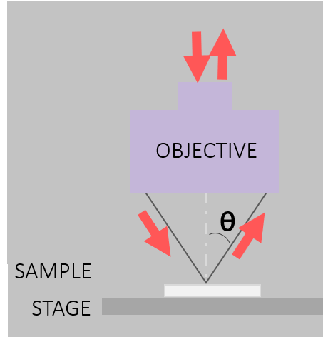
---
language: "en"
---
# Remote Access
This page describes [Remote access](https://midir.lightsource.ca/mid-infrared-spectromicroscopy-mid-ir/remote-access.md#About-Remote-Access-at-Mid-IR) for the Mid-IR beamline. Remote connection to our endstations may be used in addition to on-site user access. [Contact](https://midir.lightsource.ca/mid-infrared-spectromicroscopy-mid-ir/contact.md) Mid-IR staff to discuss the access mode that will suit your experiment best.
## About Remote Access at Mid-IR
For remote access at Mid-IR, the user designs the experiment, [++mails++](https://midir.lightsource.ca/mid-infrared-spectromicroscopy-mid-ir/shipping.md) samples, controls the instrument and analyzes the data. The beamline staff assist with connecting to the instrument computer and mounting/changing samples. They will be your primary contact for any on-site activity.
It is the user's responsibility to ensure the [++samples++](https://midir.lightsource.ca/mid-infrared-spectromicroscopy-mid-ir/samples.md) are appropriate for the desired measurements and are labelled appropriately. Measurement parameters and sample priority should be determined before beam time with the assistance of the beamline staff.
### Experiment Planning
Decide the sample holder requirements for your sample: on a window; in a liquid cell, etc (more examples on [++Samples++](https://midir.lightsource.ca/mid-infrared-spectromicroscopy-mid-ir/samples.md) page) and outline how your sample should be mounted in instructions for beamline staff.
Have your samples organized in order of priority. Communicate with beamline staff about how your beam time will be organized and how often you will require samples to be changed.
### Shipping samples
Please see details in [++Shipping++](https://midir.lightsource.ca/mid-infrared-spectromicroscopy-mid-ir/shipping.md) section on how to mail your samples to Mid-IR.
Samples can be shipped refrigerated or frozen if necessary and it is possible to [++store++](https://midir.lightsource.ca/mid-infrared-spectromicroscopy-mid-ir/samples.md#Storage) them in the fridge or the freezer when they arrive. If the measurement timing of sample is time sensitive, please make sure beamline staff know!
### Communication during your beam time
Beamline staff will work with you during your beam time to set up the experiment, change samples and help out should you run into any problems. There are many options for communication. Please make a plan with beamline staff before your beam time for communication method.
1. Microsoft Teams (preferred)
2. email
3. phone call/text
4. other, specify.
### Controlling the instrument
We use [NoMachine](https://midir.lightsource.ca/mid-infrared-spectromicroscopy-mid-ir/nomachine.md) to connect to the Mid-IR beamline endstation computers. Set-up and connection details can be found on the [NoMachine](https://midir.lightsource.ca/mid-infrared-spectromicroscopy-mid-ir/nomachine.md) page. Remote controlling the instrument is similar, but different than being on-site with the instrument. At the beginning of your beam time you will be given an orientation of instrument operations. It is recommend you have a good internet connection and an external monitor (if connecting with a notebook computer).
### Data transfer and analysis
At the end of your beam time, you will be contacted that your experimental run is complete and will be given a timeline on when to expect your data. Data can be transferred to your endpoint through [Globus](https://midir.lightsource.ca/mid-infrared-spectromicroscopy-mid-ir/data-transfer.md). Alternatively, the user can send a link to an uploading virtual drive such as OneDrive or send USB drives or Hard drives and the data can be mailed to the user.
For the general user program, data analysis is the responsibility of the user, but beamline staff are here to help! If you don't know where to start, please read the [++data analysis++](https://midir.lightsource.ca/mid-infrared-spectromicroscopy-mid-ir/software.md) user guide, watch [++tutorials++](https://midir.lightsource.ca/mid-infrared-spectromicroscopy-mid-ir/tutorials.md) and test out sample [++workflows++](https://midir.lightsource.ca/mid-infrared-spectromicroscopy-mid-ir/workflows.md). And of course, don't hesitate to [++contact us++](https://midir.lightsource.ca/mid-infrared-spectromicroscopy-mid-ir/contact.md) with your questions.
### Data analysis computer
If you are collecting hyperspectral images, it is likely your personal computer will not be able to handle the quantity of data. Please [talk to beamline staff](https://midir.lightsource.ca/mid-infrared-spectromicroscopy-mid-ir/contact.md) about using NoMachine to connect to the data analysis computer.
---
language: "en"
---
# Research Highlights
REsearch Highlights
## FTIR Proves Effectiveness of a New and Improved Method for Cleaning Eggs
### Published: February, 2025
| Before hitting the shelves at your local grocery store, eggs roll through decontamination routines such as washing with hot water and drying - but sometimes these conventional methods damage egg quality. Researchers from the University of Saskatchewan use our [++Agilent++](https://midir.lightsource.ca/mid-infrared-spectromicroscopy-mid-ir/agilent.md) endstation and data collected with [++ATR-FTIR++](https://midir.lightsource.ca/mid-infrared-spectromicroscopy-mid-ir/attenuated-total-reflection.md) to investigate new eggshell washing practices. Additional Synchrotron X-ray micro-Computed Tomography (micro-CT) data was collected using the BioMedical Imaging and Therapy ([++BMIT++](https://bmit.lightsource.ca/)) beamline! In combination, these results prove the effectiveness of cold plasma, a non-thermal, chemical-free and eco-friendly technique for the decontamination of eggshells without sacrificing egg protein quality or eggshell cuticle coverage. **Read the paper!** Movasaghi, M.; Heydari, M.M.; Schwean-Lardner, K.; Kirychuk, S.; Thompson, B.; Zhang, L. Investigating cold plasma jet effectiveness for eggshell surface decontamination. *Food Control* **2025** , *168* , 110928. DOI: [++https://doi.org/10.1016/j.foodcont.2024.110928++](https://doi.org/10.1016/j.foodcont.2024.110928) |  |
|---------------------------------------------------------------------------------------------------------------------------------------------------------------------------------------------------------------------------------------------------------------------------------------------------------------------------------------------------------------------------------------------------------------------------------------------------------------------------------------------------------------------------------------------------------------------------------------------------------------------------------------------------------------------------------------------------------------------------------------------------------------------------------------------------------------------------------------------------------------------------------------------------------------------------------------------------------------------------------------------------------------------------------------------------------------------------------------------------------------------------------------------------------------------------------------------------------------------------------------------------------------------------------------------|-------------------------------------------------------------------------------------------------------------------------------------------------------------------------------|
Research Highlights
## THE FATE OF BIO-PLASTICS
### FTIR Mapping compares conventional and biobased plastic products in the marine biodegredation pathway.
#### Published: July, 2024
Plastic-based and bio-plastic disposable materials inevitably become contaminants in our ocean ecosystems. Plastics will biodegrade and microbial ecosystems will begin to colonize them. Researchers from Memorial University in St. John's Newfoundland and Laborador use the [++Agilent endstation++](https://midir.lightsource.ca/mid-infrared-spectromicroscopy-mid-ir/agilent.md) at the Mid-IR beamline to image petroleum- and bio-based plastics. The biochemical impacts of the materials are shown, providing insights into their enviornmental impacts and helping to inform recycling policy changes.
**Read the paper!**
Cao, Yiqi; Zhang, Baiyu; Song, Xing; Dong, Guihua; Zhang, Yuanmei; Chen, Bing. Polyhydroxybutyrate Plastics Show Rapid Disintegration and More Straightforward Biogeochemical Impacts than Polyethylene under Marine Biofragmentation. *Environ Sci Technol* **2024** ,*58* (32). DOI:
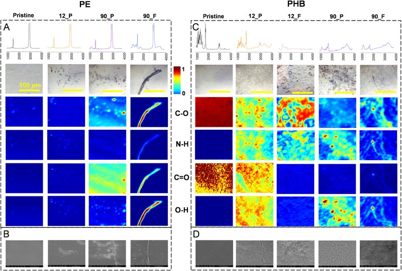
*** ** * ** ***
Research Highlights
## PINPOINTING CLUBROOT RESISTANCE IN CANOLA
### FTIR Microscopy shows importance of cell wall structure in clubroot resistance
#### Published: July 22, 2024
Clubroot is of serious concern to the agricultural community. Researchers from the University of Saskatchewan and Agriculture and Agri-Food Canada use the [++Agilent endstation++](https://midir.lightsource.ca/mid-infrared-spectromicroscopy-mid-ir/agilent.md) at the Mid-IR beamline to compare susceptible and resistant varieties of canola roots which have been innoculated with clubroot. These researchers use [++FTIR microspectroscopy mapping++](https://midir.lightsource.ca/mid-infrared-spectromicroscopy-mid-ir/focal-plane-array.md) to better understand the innoculation pathway and identify key genes expressed in the cell walls of resistant varieties of canola.
**Read the paper!**
Tu, J.; Li Qin; Karunakaran, C.; Wei, Y.; Peng, G.. Lignin accumulation in cell wall plays a role in clubroot resistance. *Front. Plant Sci.,* **2024** ,*15* .
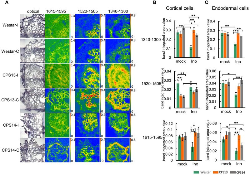
*** ** * ** ***
Research Highlights
## INVESTIGATING SOLID-STATE BATTERIES
### hATR Microscope Reveals Post-Cycling Changes of Advanced Electrolyte Materials
#### Published: June 12, 2024
Researchers from the University of Alberta and the University of British Columbia use the horizontal attenuated total reflection (hATR) microscope of the Mid-IR beamline to study solid-state batteries. They made use of our focal plane array (FPA) detector to track heterogeneities in the electrolyte surface during changing/discharging.
**Read the paper!**
Dong, S.; Xie, G.; Xu, S.; Tan, X.; Chaudhary, M.; Zhang, Y.; Wu, R.; Wen, F.; Ayranci, C.; Michaelis, V.K.; Quirk, A.; Rosendahl, S.M.; Liu, J.; Fleischauer, M.D.; Sang, L. Cellulose-Encapsulated Composite Electrolyte Design: Toward Chemically and mechanically enhanced Solid-Sodium Batteries. *ACS Nano* **2024** ,*18* (25), 16285-16296.
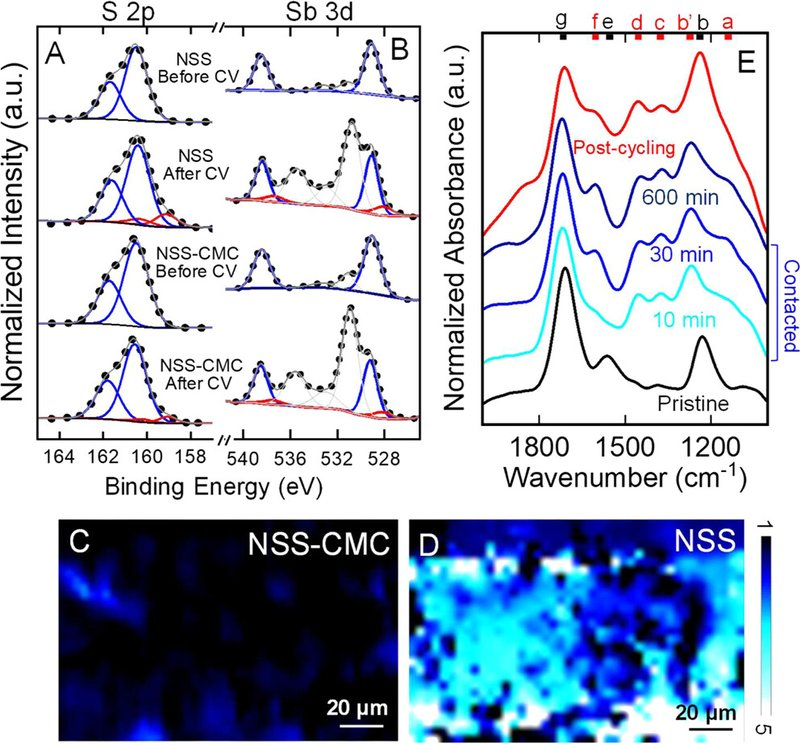
*** ** * ** ***
Research Highlights
## OIL SPILL CLEANUP SOLUTIONS
### Synchrotron FTIR Identifies Performance and Composition of Oil-Remediating Aerogels
#### May 1, 2024
Researchers from Memorial University in St. John's Newfoundland and Laborador use the [++Bruker endstation++](https://midir.lightsource.ca/mid-infrared-spectromicroscopy-mid-ir/bruker.md) at the Mid-IR beamline to analyze an oil-absorbing aerogel. The chemical composition of the aerogel was identified with SR-FTIR. Furthermore, the aerogel was imaged before and after UV-irradiation to identify UV-induced changes in the material.
**Read the paper!**
Wang, H.; Chen, X.; Chen, B.; Zhao, Y.; Zhang, B. Development of a spiropyran-assisted cellulose aerogel with switchable wettability as oil sorbent for oil spill cleanup. *Sci Total Env* **2024** ,*923* , 171451. [++https://doi.org/10.1016/j.scitotenv.2024.171451++](https://doi.org/10.1016/j.scitotenv.2024.171451).
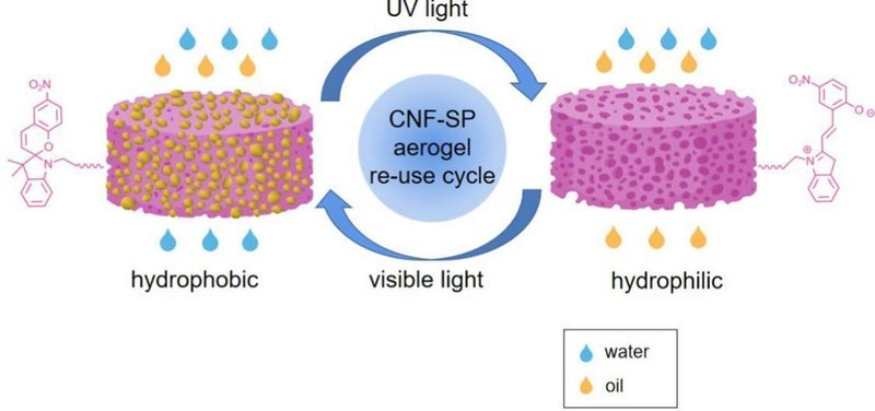
*** ** * ** ***
Research Highlights
## THE LIFE OF BIOCHAR
### SR-FTIR Informs Environmental Impacts of Fertilizer Use
#### Published: March 3, 2024
Biochar is a form of charcoal - formed from pyrolysis of plant material. This carbon-rich material is a key player in soil health - benefiting both nutrient cycling and microorganisms. However, biochar breakdown processes - and its affinity to bind with environmental pollutants - make the long-term utilization of this fertilizer an environmental risk. Synchrotron FTIR analysis using the [++Bruker++](https://midir.lightsource.ca/mid-infrared-spectromicroscopy-mid-ir/bruker.md) endstation analyzed surfaces of biochar, identifying functional group changes with typical uses of biochar and its interractions with pollutants. This study contributes to understanding the aging and contaminant distribution processes of biochar in the natural environment.
**Read the paper!**
Shen, J.; Huang, G.; Yao, Y.; Zhang, P.; Rosendahl, S. Surface Alteration on Biochar in Long-Term Application: Insights into Pyrolysis, Freeze-Thaw Aging, and Dissipation. *Surf. Interfaces* **2024** , 104118. DOI: [++10.1016/j.surfin.2024.104118++](https://doi.org/10.1016/j.surfin.2024.104118).
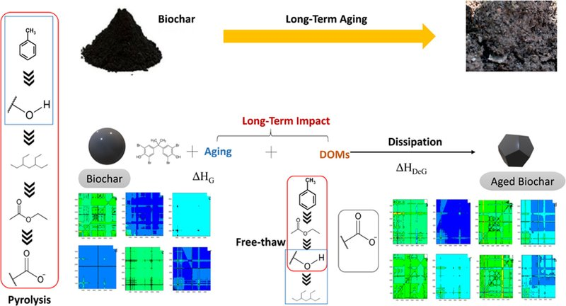
*** ** * ** ***
Research Highlights
## UNDERSTANDING THE POST-STROKE BRAIN
### Imaging the Neurovascular Unit with FTIR
#### Feb. 14, 2024
This eview article advertises the use of [infrared chemical imaging](https://midir.lightsource.ca/mid-infrared-spectromicroscopy-mid-ir/focal-plane-array.md) conducted with the [++Agilent microscope++](https://midir.lightsource.ca/mid-infrared-spectromicroscopy-mid-ir/agilent.md) at the Mid-IR beamline to identify key biomarkers of stroke within brain tissue. Researchers use FTIR imaging to analyze changes in brain tissue enabling a more holistic approach to characterizing pathophysiological responses - helping us understand the processes occuring in stroke incidence and identifying risk factors - in disease and disease models. A detailed description of machine learning with [++Orange/Quasar data analysis++](https://midir.lightsource.ca/mid-infrared-spectromicroscopy-mid-ir/software.md) is presented to aid differentiation and characterization of tissue sections. [Tissue preparation](https://midir.lightsource.ca/mid-infrared-spectromicroscopy-mid-ir/samples.md) methods suitable for FTIR imaging analysis are also described.
**Read the paper!**
Boseley, R.E.; Sylvain, N.J.; Peeling, L.; Kelly, M.E.; Pushie, M.J. A review of concepts and methods for FTIR imaging of biomarker changes in the post-stroke brain. *Biochim Biophys Acta Biomembr* **2024** , *1866* (3), 184287. [++https://doi.org/10.1016/j.bbamem.2024.184287++](https://doi.org/10.1016/j.bbamem.2024.184287)
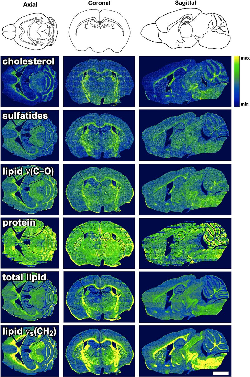
*** ** * ** ***
Research Highlights
## IDENTIFYING RISKS OF PLASTIC POLLUTANTS
### Synchrotron FTIR Reveals Nanoplastic Toxicity to Microalgae
#### Oct. 1, 2023
Synchrotron FTIR spectroscopy using the [++Bruker endstation++](https://midir.lightsource.ca/mid-infrared-spectromicroscopy-mid-ir/bruker.md) at the Mid-IR beamline reveals changes in microalgal lipids and proteins indicating nanoplastic toxicity and risk assessment of emerging pollutants. Researchers conclude the mechanism of toxicity induced by nanoplastics to microalgae under multiple environmental conditions - contributing to emphasize the importance of ecological risk assessments and environmental management of nanoplastics.
**Read the paper!**
Gao, S.; Huang, G.; Zhang, P.; Xin, X.; Yin, J.; Han, D.; Rosendahl, S.; Read, S. Toxicity and mechanism of nanoplastics to phytoplankton in high-latitude aquatic ecosystems of Canadian prairie: Effects of multiple environmental factors. *Sci. Total Environ* **2023** , 893, 164676. [++https://doi.org/10.1016/j.scitotenv.2023.164676++](https://doi.org/10.1016/j.scitotenv.2023.164676).
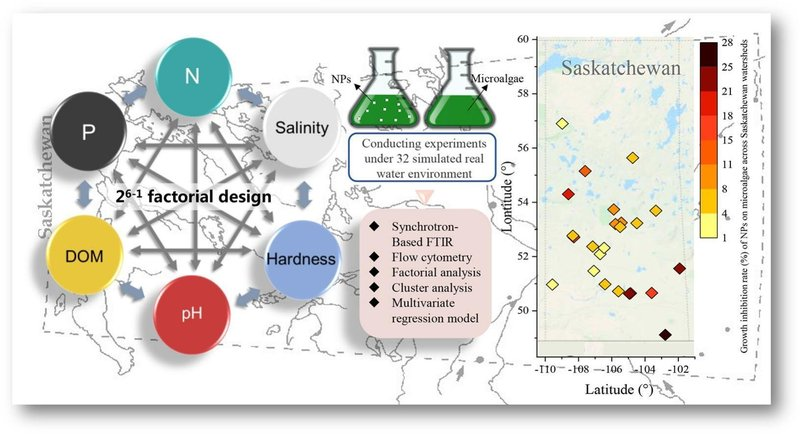
*** ** * ** ***
Research Highlights
## DEVELOPING NUTRITIOUS AND DELICIOUS PLANT-BASED PROTEIN
### FTIR Imaging Characterizes Secondary Protein Structures
#### June 1, 2023
Plant-based meat alternatives are increasingly in demand. Innovative strategies are required to provide consumers with appropriately textured and nutrient-dense protein sources. This study aims to increase the nutritional value of the food, reduce production costs, and improve the environmental footprint of plant-based meat analogues. Infrared imaging using the [++Agilent microscope++](https://midir.lightsource.ca/mid-infrared-spectromicroscopy-mid-ir/agilent.md) at the Mid-IR beamline was used to analyze plant protein secondary structures. [++Orange/Quasar data analysis++](https://midir.lightsource.ca/mid-infrared-spectromicroscopy-mid-ir/software.md) using K-means clustering was used to identify unique patterns in the data.
This publication represents cross-beamline collaboration. The FTIR results complement synchrotron-based micro-computed tomography collected at [++BMIT++](https://bmit.lightsource.ca/).
**Read the paper!**
Dobson, S.; Stobbs, J.; Laredo, T.; Marangoni, A.G. A facile strategy for plant protein fiber formation without extrusion or shear processing. *Innov Food Sci Emerg Technol* **2023** , 86, 103385. [++https://doi.org/10.1016/j.ifset.2023.103385++](https://doi.org/10.1016/j.ifset.2023.103385)
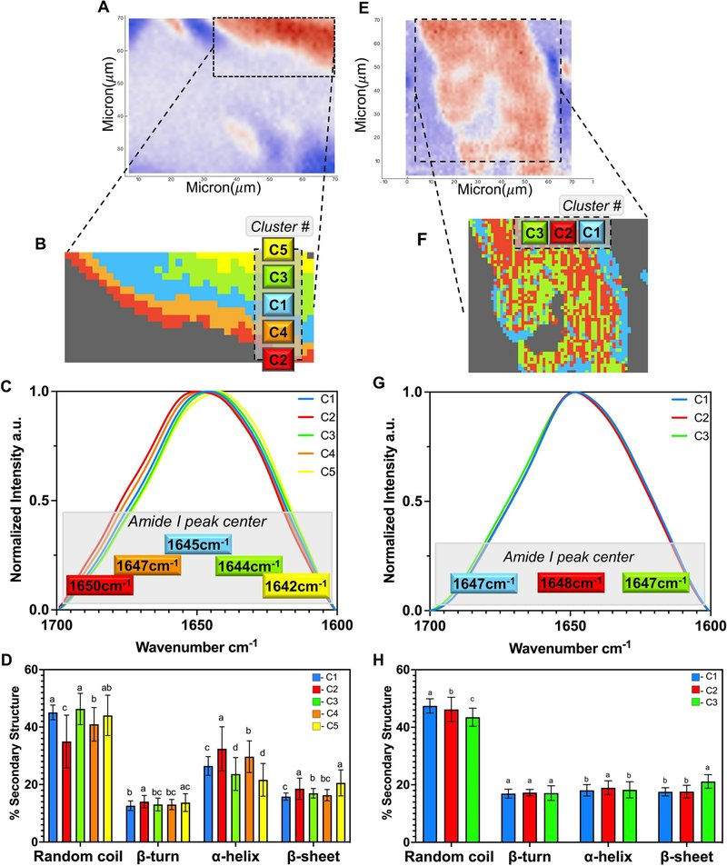
*** ** * ** ***
Research Highlights
## ADVANCING PIPE MANUFACTURING
### IR Imaging Coupled with AI Identifies Aging in Pipes
#### April 25, 2023
Learned representations of hyperspectral infrared images obtained at the Mid-IR beamline are used to extract physicochemical spectral markers of aging and cracking in cross-linked polyethylene pipes. Indicators of degredation were identified and learned by the model and applied to identify both unused and damaged pipes.
These results will help pipe manufacturers optimize fabrication methods and improve stress-testing protocols, extending the use of their pipes into ever more vigorous and challenging operating environments. The study also highlights the use of representation learning via deep generative modeling to provide interpretable information from complicated data sets - unlocking new capabilities for hyperspectral IR imaging using the [++Agilent microscope++](https://midir.lightsource.ca/mid-infrared-spectromicroscopy-mid-ir/agilent.md) at the Mid-IR beamline to solve complex problems across global sectors.
**Read the paper!**
Grossutti, M.; D'Amico, J.; Quintal, J.; MacFarlane, H.W.; Wareham, C.; Quirk, A.; Dutcher, J.R. *ACS Appl Mater Interfaces* **2023** , *15* (18), 22532-22542.
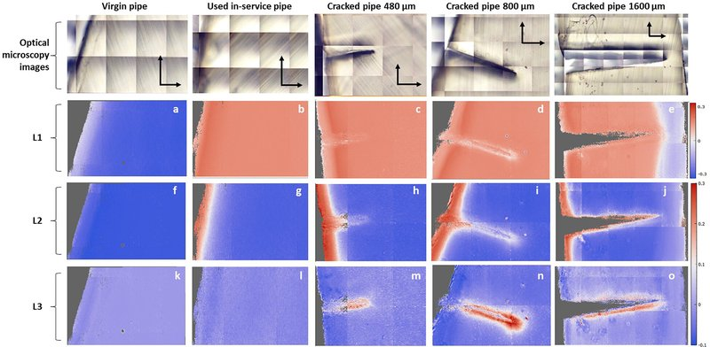
*** ** * ** ***
Research Highlights
## IMPROVING NUTRITION OF CANADIAN GRAINS
### FTIR Analyzes Pea and Lentil Flour Processing Effects
#### Feb. 2, 2023
Pulse production is an integral pillar of the Canadian agricultural economy. Changes to the nutritional properties and protein structure of green lentil and yellow pea flour post-seed milling and processing are determined. The authors used the [++Agilent endstation++](https://midir.lightsource.ca/mid-infrared-spectromicroscopy-mid-ir/agilent.md) at the Mid-IR beamline to examine pulse flours - reporting changes to protein structure and an improvement in starch digestibility with tempering and IR heating. Sample preparation used KBr pellets / flour mixtures.
**Read the paper!**
Laing, E.; Stone, A.K.; Shi, D.; Pickard, M.; House, J.D.; Wang, N.; Nickerson, M.T. Effect of infrared heating on the nutritional properties of yellow pea and green lentil flours. *Cereal Chem* **2023** , *100* , 614-627.

*** ** * ** ***
Research Highlights
## TIME-RESOLVED SPECTROELECTROCHEMISTRY WITH IRSWEEP
### Dual comb IR spectroscopy for time resolved spectroscopy
#### Published: Oct. 21, 2020
A dual infrared frequency comb spectrometer with heterodyne detection has been used to perform time-resolved electrochemical attenuated total reflectance surface-enhanced infrared absorption spectroscopy (ATR-SEIRAS). In collaboration with the [++**Canadian Light Source**++](https://www.lightsource.ca/facilities/beamlines/cls/beamlines/mid-ir.php) and [++**IRsweep**++](https://irsweep.com/), the desorption of a monolayer of a pyridine derivative with 10 μs time resolution and a detection limit of 5% of a monolayer was demonstrated. The applications of the method are potentially immense for the study of short lived adsorbed species during electrocatalysis. You can access the [++**pre-print**++](https://harvest.usask.ca/handle/10388/12909) ( ) in the UofS open access repository.
[++**Analytical Chemistry**++](https://pubs.acs.org/doi/10.1021/acs.analchem.0c00260).( ).
**Read the Paper!**
Lins, E.; Read, S.; Unni, B.; Rosendahl, S.M.; Burgess, I.J. Microsecond Resolved Infrared Spectroelectrochemistry Using Dual Frequency Comb IR Lasers. *Anal. Chem.* **2020** , 92(*9* ), 6241--6244. DOI: .
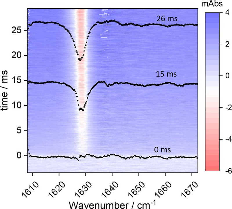
*** ** * ** ***
Research Highlights
## STUDYING LIVING CELLS
### Synchrotron FTIR spectromicroscopy as a tool for studying populations and individual living cells of green algae
#### Published: Oct. 21, 2020
Fourier transform infrared (FTIR) spectromicroscopy was used to study individual living cells of three closely-related species of the green algal genus Chlamydomonas. Three types of spectral variation were observed between individual cells within a single culture, as well as between different cultures: variation around a mean, individual outliers, and the presence of subpopulations. By understanding and controlling this variation, we were able to spectroscopically differentiate between the three closely-related species. Spectral differences were confirmed using principal component analysis, leading to an understanding of the biochemical differences between species. This work highlights the additional information obtained by studying individual cells, and has implications for more traditional bulk measurements.
Read the Paper!
Goff, K.L.; Ellis, T.H.; Wilson, K.E. Synchrotron FTIR spectromicroscopy as a tool for studying populations and individual living cells of green algae. *Analyst* **2020**, 24. DOI: 10.1039/d0an01386b.
*** ** * ** ***
## Research Highlights
## RECENT PUBLICATIONS
### We would love to celebrate your accomplishments!
#### Sept. 23, 2020
Yay data! Publishing research articles? Submitting a thesis? Attending conferences? Send us an [++email++](https://midir.lightsource.ca/mid-infrared-spectromicroscopy-mid-ir/contact.md)! Include a figure and summary of the work and we'll highlight it in our research news.
Make sure you [++report your published works++](https://www.lightsource.ca/users/your-cls-experiment/after-your-beamtime.php#ReportingPublications) to the CLS, too!

---
language: "en"
---
# Sample Environments
We see a plethora of sample types and sampling environments at the Mid-IR beamline spanning research sectors.
Each of our endstations provide dry air purge to remove infrared-absorbers from the sampling enviornments such as gas-phase water and atmospheric CO~2~. If you're bringing custom sampling environments, have a chat with beamline staff about how we can customize the dry-air purge environment to be suitable for your set-up.
A variety of custom sample environments can be situated onto our microscopes, and we have custom optics available open to new developments to make your experiments work on our beamline. [Contact](https://midir.lightsource.ca/mid-infrared-spectromicroscopy-mid-ir/contact.md) beamline staff to discuss and take a look at the [sample preparation tools and equipment](https://www.lightsource.ca/facilities/laboratories-equipment.php) available for booking during your experiment.
Mention any custom equipment and tools you'd like to bring to the beamline in your proposal as they will likely require review and approval before they can be brought on-site.
We have a temperature controlled stage which can be used to keep samples at a specific temperature and for *in-situ* temperature experiments. Set-up a meeting with us to talk more about how the [Linkam FTIR600 stage](https://canadianlightsource.atlassian.net/wiki/display/MISM/User+Guide%3A+Sample+Heating+%26+Cooling+with+Linkam+Stage) can work for your experiment.
Here's more information about preparing [Samples](https://midir.lightsource.ca/mid-infrared-spectromicroscopy-mid-ir/samples.md) for beamtime!
---
language: "en"
---
# Samples
## Sample Types
The Mid-IR beamline is ideal for studying biomedical and biological materials, agricultural and environmental samples, materials such as polymers, art specimens, and much more!
This guide is designed as a starting point for sample preparation for experiments at Mid-IR. Please talk to beamline staff about your specific samples and unique requirements to ensure high quality data collection is possible.
### Resources
Mid-IR beamline staff are a great point of contact to discuss best practices for preparing and storing your samples for our beamline. [Contact us](https://midir.lightsource.ca/mid-infrared-spectromicroscopy-mid-ir/contact.md) to discuss!
Check out our [**Sample preparation and considerations**](https://midir.lightsource.ca/mid-infrared-spectromicroscopy-mid-ir/reading.md#Sample-preparation-and-considerations) section on our Reading page for more in-depth information.
## Sample Preparation
The Canadian Light Source has several user accessible labs. Basic lab access requires the completion of CLS Laboratory Safety Training. General lab equipment includes: Ultrasonic cleaners, ovens, balances, pH meters, pipettors, microscope, fume hoods, hot plate, freezers, refrigerators, and much more! Specialized equipment are in the electrochemistry lab, SyLMAND, earth sciences lab and life sciences lab.
Our [**CLS Laboratories and Equipment**](https://www.lightsource.ca/facilities/laboratories-equipment.php) page contains full details and locations of each lab.
## Sample Holders
There are many standard sample holders available to use at Mid-IR
### Transmission Holders
A) holder for two 25 mm CaF~2~ windows
B) Liquid cell, uses 25 mm CaF~2~ windows
C) Holder for one 25 mm CaF~2~ window
D) Holder for four 10 mm CaF~2~ windows
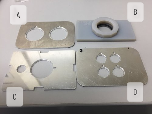
## Windows
### About windows
Calcium Fluoride (CaF~2~) are commonly used at Mid-IR as a sample support. The windows come in a variety of sizes and thicknesses. Sizes 25 mm and 10 mm (thickness 1 mm) are supported with the standard sample holders, other sizes can be accommodated, but arrangements must be made well in advance of beam time. Properties of mid-IR optic materials are listed in the table below.
|---|---|---|---|---|
| **Comparison of Common Infrared Optic Materials** |||||
### Where to buy
User groups will supply their own IR windows. They can be purchased from [Crystran](https://www.crystran.co.uk/windows).
### How to clean
Windows of different materials will have different cleaning protocols.
CaF~2~ windows can be cleaned with water and are acid/base resistant. The windows can be dried and gently polished with lens paper
BaF~2~ windows are extremely sensitive to water and should not come into contact with water.
\*\* Using clean windows is incredibly important for collecting high quality data \*\*
## Storage
Samples mailed to the Mid-IR for mail-in or remote access experiments can be stored until beamtime. Some options include
* Refrigerator (5 **°**C)
* Freezer (-20 **°**C)
* Extra cold freezer (-80 **°**C)
* Desiccator
Need a storage location not listed? Please [++contact++](https://midir.lightsource.ca/mid-infrared-spectromicroscopy-mid-ir/contact.md) beamline staff to figure out a solution. Check out the [CLS Laboratories \& Equipment](https://www.lightsource.ca/facilities/laboratories-equipment.php) page for storage locations.
## Sample disposal
Samples need to be removed when your beam time is finished. In the case of mail-in and remote access, samples will be returned to the user or disposed of if specific instructions are provided.
---
language: "en"
---
# Shipping
## CLS Guidelines
### [++Shipping++](https://www.lightsource.ca/users/your-cls-experiment/practical-information/shipping.php) to the Canadian Light Source
* Ship one week before your scheduled experimental collection time
* Users are responsible for the cost of transportation and customs clearance for shipments; C.O.D. packages will not be accepted
* All packages must be addressed to a permanent staff member of the CLS, preferable to the beamline scientist or associate scientist you have been working with
* To expedite outgoing shipments, you can make round-trip shipping arrangements from your home institution. Please advise Shipping \& Receiving if you have made these arrangements
#### Address Package as follows:
**c/o Dr. Scott Rosendahl** (or applicable recipient)
**Mid Infrared Spectromicroscopy, Project Number**
**Canadian Light Source, Inc.**
**University of Saskatchewan**
**44 Innovation Boulevard**
**Saskatoon, SK**
**Canada S7N 2V3**
\*\*\*Please notify [++CLSI Receiving++](https://www.lightsource.ca/users/your-cls-experiment/practical-information/shipping.php#GeneralInformation) at least 1 week prior to arrival to advise of any shipments that will arrive before or during your beamtime. This is especially important if you intend to ship hazardous materials where storage of the shipment may require advance planning; if there are any special handling instructions.\*\*\*
### Some notes on shipping
* Shipping frozen of refrigerated samples please label appropriately and notify beamline staff
* If you are sending a hard drive or thumb drive, please label it with the principal investigator's name, enclose in protective wrapping, and include in shipping container.
* [Contact](https://midir.lightsource.ca/mid-infrared-spectromicroscopy-mid-ir/contact.md) Mid-IR staff with specific sample handling requirements before your samples arrive at the CLS
* **Packing:** Whenever possible, materials should be shipped in reusable containers (i.e. cardboard boxes or wood crates). These containers will be used to repackage items for return to your location. It is recommended that you include a packing slip with the destination address inside the package in case of lost or damaged shipments.
## Return Shipping
Please complete [++Shipping Order Form++](https://www.lightsource.ca/users/your-cls-experiment/practical-information/user-shipping-form.pdf)
The most important information required is:
* Your FedEx account number.*\["Consignee's Courier Account #" field\]*
* The correct return address.
* Shipping speed *(optional).*
* **\*Overnight** shipping is fast and recommended if your samples need specific storage conditions
* **Ground/economy** shipping costs significantly less but may take a week or more so your samples should be stable
* **2-day** shipping might be a good compromise.
* **\*If nothing is indicated, overnight shipping will be assumed.** Please allow sufficient time for outgoing shipments.
## Shipping Hazardous Materials
Users should consult with a Materials Specialist at their location for the proper handling and shipping of hazardous materials.
Hazardous materials shipped to or from the CLS by truck must be packaged and handled according to Transportation of Dangerous Goods (TDG) regulations. Hazardous materials shipped by air must meet International Air Transportation Association (IATA) regulations.
Each shipment of hazardous materials must include pertinent Materials Safety Data Sheets (MSDS). Two copies of MSDS are required for products covered under WHIMIS along with appropriate warning labels.
Ship the smallest quantity necessary for your experiment.
## Contact Information
### [++Shipping Contact++](https://www.lightsource.ca/users/your-cls-experiment/practical-information/shipping.php#GeneralInformation)
Location: Room 1055, CLS Main Floor Level
Hours: Monday - Friday 8:00 a.m. to 4:30 p.m.
Phone: 1-306-657-3657
Fax: 1-306-657-3533
Email: stores@lightsource.ca
---
language: "en"
---
# Software
**Quasar** is an open‑source suite of spectroscopic data‑analysis tools built on the Orange machine‑learning and visualization platform. It enables users of all experience levels to create dynamic, reproducible workflows that integrate spectral processing, hyperspectral mapping, and multivariate analysis. ([++Orange, 2013++](https://jmlr.org/papers/volume14/demsar13a/demsar13a.pdf))
If you're new to Quasar, explore our [Getting Started](https://midir.lightsource.ca/mid-infrared-spectromicroscopy-mid-ir/getting-started.md) guide, [Tutorials](https://midir.lightsource.ca/mid-infrared-spectromicroscopy-mid-ir/tutorials.md), [Tips and tricks](https://midir.lightsource.ca/mid-infrared-spectromicroscopy-mid-ir/tips-and-tricks.md) and example [Workflows](https://midir.lightsource.ca/mid-infrared-spectromicroscopy-mid-ir/workflows.md) to begin working with FTIR data.
Quasar offers fast, flexible spectral‑processing tools---baseline subtraction, normalization, FFT, EMSC, peak analysis, differentiation, smoothing, and more. These features can be combined with sample metadata for multivariate methods such as regression, classification, clustering, PCA, and hierarchical analysis. Workflows can be shared to ensure consistent, reproducible analysis across projects.
Advanced users can integrate custom Python [code](https://midir.lightsource.ca/mid-infrared-spectromicroscopy-mid-ir/custom-code.md) or [figures](https://midir.lightsource.ca/mid-infrared-spectromicroscopy-mid-ir/custom-figures.md) directly into workflows, leveraging the broader scientific Python ecosystem. Community [contributions](https://midir.lightsource.ca/mid-infrared-spectromicroscopy-mid-ir/development.md) are welcome. [Connect](https://midir.lightsource.ca/mid-infrared-spectromicroscopy-mid-ir/contact.md) with beamline staff to learn more, request new features or share your data‑processing needs. We're here to support your analysis.
For more information and to download the software, visit [++https://quasar.codes++](https://quasar.codes/).
If you used Quasar or Orange-Spectroscopy for your data analysis, please include a citation.
For more information, see [Quasar - Publications](https://quasar.codes/publications/) and [Quasar - Citation](https://quasar.codes/citation/).
---
language: "en"
---
# Tips and tricks
This page outlines some small tips and tricks that make working with your data in Quasar even more fun and rewarding!
If you're struggling with something annoying or overly tedious, please [++let us know++](https://midir.lightsource.ca/mid-infrared-spectromicroscopy-mid-ir/contact.md).
*** ** * ** ***
## Widget Help
Each widget has help documentation!
Access it:
* Right click on widget, select help
Information in Help:
* Types of inputs (data, widgets, etc)
* Types of outputs
* Purpose of the widget
* How to use the widget
* Widget options
* References
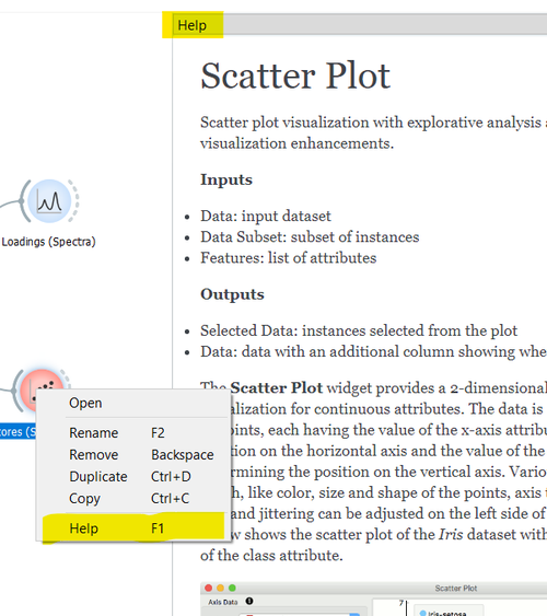
*** ** * ** ***
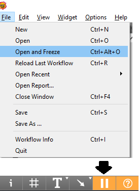
## Open and Freeze
* If you want to look at something in your workflow, but don't want to load the dataset or trigger any data processing load it with the "open and freeze" option located under the file menu! Once you have everything sorted you can click the pause button in the bottom left hand corner and processes will proceed as expected.
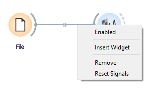
* If you have a large workflow and want to stop some parts from updating, you can disable a signal connection by right-clicking on it and deselecting "Enabled".
* Data file locations are saved relative to the workflow file, so keeping your data and workflows together makes sharing analysis easier!
*** ** * ** ***
## Data analysis not working as expected?
* One of the first things to check is the wire connections between widgets
* Double click on wire
* Pop-up with connection options opens
* Draw connection
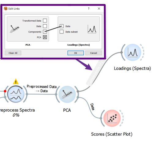
*** ** * ** ***
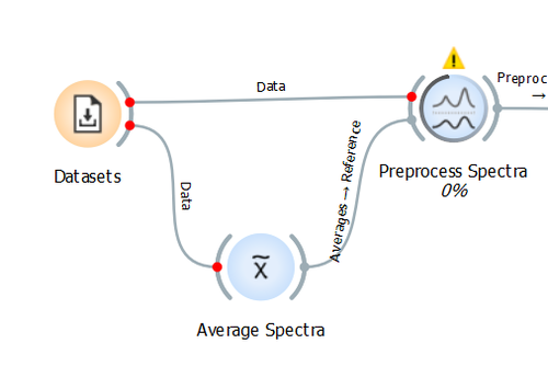
## Red dots at Widget Connections
... what does it mean?
* Data is still being loaded from the File or Datasets widgets
OR
* Data is being processed in a widget and isn't ready to be passed to the next connected widget
When the red dots disappear it's go time - the data has been passed to the widget!
*** ** * ** ***
## Hyperspectra
One of the powerful features of the [++Hyperspectra++](https://orange.biolab.si/widget-catalog/spectroscopy/hyperspectra/) widget is custom region selection.
* When selecting a region, you can use a simple rectangle or a custom polygon.
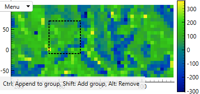
Menu → Select (rectangle)
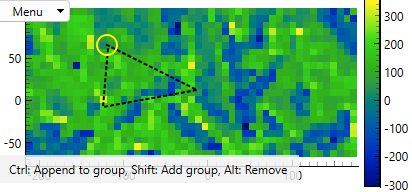
Menu → Select (polygon)
Keyboard Shorcuts
Hold CTRL to Append to group
Hold SHIFT to Add group
Hold ALT to Remove selected points
Careful: Removing regions is done all-or-nothing by clicking on the white boundary area of the figure. This can also happen accidentally as clicking a pixel without holding buttons will discard the previously selected points/regions.
* In the case of multiple region selection, you must use the "Data" output (as opposed to the Selection output) to see the annotated spectra.
* One can use the 'Edit Domain' widget to *rename* the selected regions:
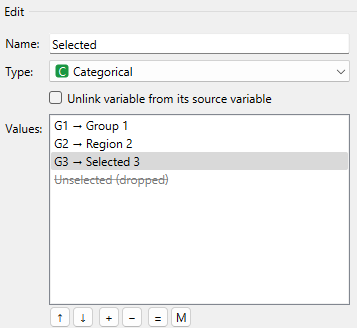
Edit Domain → Double-click to modify Values
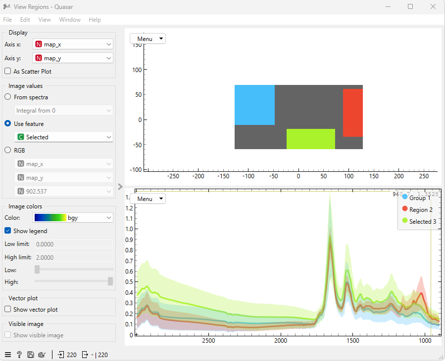
View Regions with Hyperspectra Widget
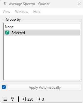
Average selected spectra by Category
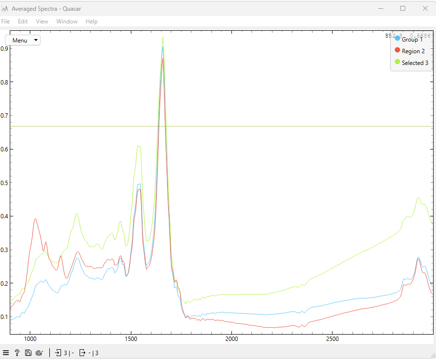
View averaged spectra with Spectra Widget
*** ** * ** ***
## Have a great tip or trick? Please send it to [++us++](https://midir.lightsource.ca/mid-infrared-spectromicroscopy-mid-ir/contact.md)!
[Next Page](https://midir.lightsource.ca/llms-full.txt/1)